Am Fam Physician. 1998;57(11):2805-2806
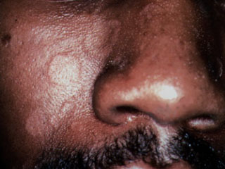
A 35-year-old black man presented to the office with a number of slightly itchy small round lesions on his face (see the accompanying photograph). The lesions were located primarily on the cheeks and the mid-parts of the face, but had also become confluent along the scalp line. The patient had used both over-the-counter clotrimazole (Lotrimin, Mycelex) and 0.5 percent hydrocortisone creams and had experienced some temporary relief. No lesions were found on the trunk or the extremities.

Question
Discussion
The answer is B: seborrheic dermatitis. “Petaloid seborrheic dermatitis” is a frequent presentation of seborrheic dermatitis in people with dark skin.1 Typically, patients present with red, scaly plaques in the eyebrows and along the melo-labial fold; however, many patients with dark skin present with polycyclic coalescing rings. These rings may be slightly pink or hypopigmented in color and usually do not show significant scale until the area is scraped for a potassium hydroxide (KOH) preparation. The etiology of seborrheic dermatitis is not clear and has been associated with the yeast Pityrosporum orbiculare, the mite Demodex folliculorum, various bacterial colonizations and skin response to the environment, such as changes in temperature, humidity and bath water.
Treatment usually includes the application of a topical corticosteroid containing 1 percent hydrocortisone cream. In refractory cases, 0.2 percent hydrocortisone valerate or 0.5 percent desonide creams may be applied twice daily. Use of stronger corticosteroids on areas other than the scalp is discouraged as it may lead to atrophy, telangiectases, perioral dermatitis and, in people with dark skin, noticeable hypopigmentation. Topical and systemic anti-fungal agents have occasionally been used with good results. Shampoo containing ketoconazole has helped many patients with seborrheic dermatitis of the scalp.2 Washing the hair and the face with prescription and over-the-counter dandruff shampoos containing selenium sulfide may also help, although washing the face with selenium sulfide more often than two or three times per week can be irritating.
The differential diagnosis for patients with seborrheic dermatitis is broad and may require biopsy to confirm the clinical presentation, especially when usual treatment measures do not work. Sarcoidosis can initially look like seborrheic dermatitis, although confluence along the scalp line and eyebrows is more typical of seborrheic dermatitis. Patients with sarcoidosis may respond to topical corticosteroids but usually require systemic therapy or very potent topical agents to clear the lesions.
Secondary syphilis often presents with rings similar to those pictured here, although the presence of lesions only on the face and not on the palms or soles would be most unusual. A negative serology test will exclude lues in more questionable cases. Tinea faciei may look identical to seborrheic dermatitis, but results of the KOH preparation should be positive and the patient should respond to topical antifungal medications. Discoid lupus erythematosus may start with flesh-colored rings, but hypopigmented, red papules and plaques would be a more common presentation. Diagnosis of discoid lupus is best determined using biopsy, since anti-nuclear antibody serologies are usually negative. Cutaneous T-cell lymphoma (called mycosis fungoides) may also include hypopigmentation; this is a relatively common presentation in young, dark-skinned patients. Again, biopsy for histopathology is necessary to confirm or exclude the diagnosis.