Am Fam Physician. 1998;58(2):489-490
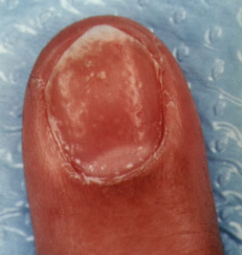
A 36-year-old man presented to a physician's office concerned about increasingly unsightly nails on both his hands and feet. Some of the nails looked worse than others, but all showed some degree of pitting, brownish discoloration of the nail bed and separation at the distal nail plate (see the accompanying photograph). He had previously been prescribed oral griseofulvin (Grisactin), in a dosage of 500 mg twice daily for one year, followed by pulse itraconazole (Sporanox), in a dosage of 200 mg twice daily for one week each month over a four-month period, without noticeable benefit. He had no other medical problems except for scaly red plaques on his knees and elbows for the past 10 years.

Question
Discussion
The answer is A: psoriatic nails. Nail changes in patients with psoriasis are manifold and may be severe enough to be confused with onychomycosis.1 Characteristic findings of psoriatic nails are pits in the nail plate and brownish discolorations of the nail bed known as oil spots (seen distally in this case). In time, nails may become thickened and dystrophic. Distal onycholysis (defined as separation of the distal nail plate) is also common and may provide a place of entry for fungal infections. It was previously thought that psoriatic nails were unlikely to become infected; however, recent studies suggest that they may be more prone to fungal superinfection than normal nails. Before the patient is treated with systemic agents, the fungal infection should be confirmed by potassium hydroxide preparation, nail plate biopsy or culture of subungual debris.
While a number of conditions can cause pits in the nail plate, the only other diagnosis to be considered in this patient would be “alopecia areata of the nails,” in which, for unknown reasons, the nail plate is covered with innumerable pits in a checkerboard pattern. Treatment of psoriatic nails is problematic because the most effective therapies have significant drawbacks. Injections of fluorouracil and corticosteroids into the matrix of the nail have shown benefit but are painful and the effect is temporary; the injections are best performed by someone experienced in this technique. Topical fluorouracil solution (Adrucil) and superpotent corticosteroids may benefit some patients. The use of occlusion boosts the efficacy of topical fluorouracil solution but increases the potential for inflammation. Occlusion also increases the risk of steroid atrophy when corticosteroids are used.
The most effective therapies are systemic agents such as methotrexate (Rheumatrex), acitretin (Soriatane) and cyclosporine (Sandimmune), but these agents would not usually be indicated for treatment of nail problems alone because of their cost and potential for systemic complications.
The nail plate contains almost no calcium and taking supplemental calcium will not “strengthen” the nails. Likewise, gelatin supplements will benefit the patient's overall nutrition more than the strength and appearance of the nails. Biotin, taken in dosages of 500 to 1,000 mg daily, has been shown to decrease nail brittleness and splitting of the leading edge in some patients.2 It must be taken for three to six months before its effect can be accurately assessed. Allergic contact dermatitis to artificial nails or to the glues used to apply them may cause pits in the nail plate but are more likely to cause distal onycholysis and Beau's lines if the inflammation is intense enough to stress the matrix and the developing nail.