Am Fam Physician. 2016;93(1):57-58
Author disclosure: No relevant financial affiliations.
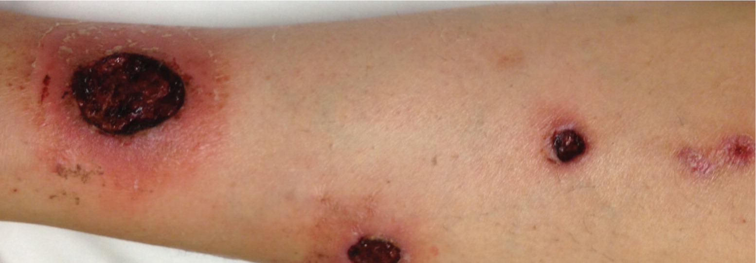
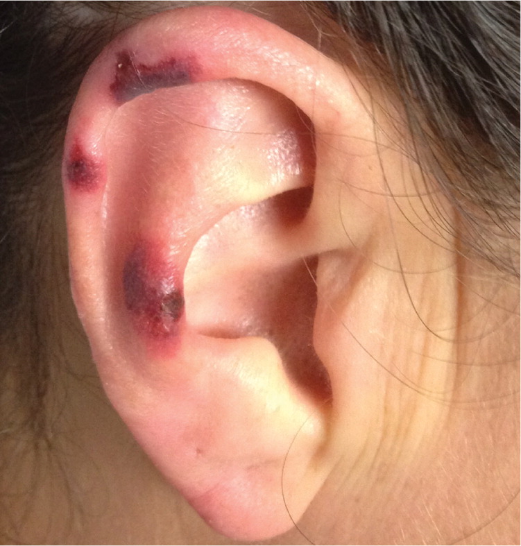
A 41-year-old woman presented to the emergency department with leg ulcers and ecchymoses on her ears that began to develop one month prior. The leg ulcers began as painful, fluid-filled blisters and evolved into ulcers with a black crust. The lesions had appeared and resolved several times over the previous three years, but she did not seek medical attention. She took prednisone intermittently for psoriasis and had a long history of cocaine abuse.
Physical examination showed multiple ulcerated lesions on the anterior aspect of both lower extremities (Figure 1) and multiple tender purpuric lesions on both ears (Figure 2). The examination showed psoriatic plaques on her legs, elbows, and fingers. Her vital signs were normal. A complete blood count, comprehensive metabolic panel, and coagulation laboratory test results were normal. Urine toxicology testing was positive for cocaine. She had an elevated C-reactive protein level (96.3 mg per L [917.16 nmol per L]) and erythrocyte sedimentation rate (48 mm per hour).


Question
Discussion
The answer is A: levamisole-induced vasculitis. A skin biopsy showed a leukocytoclastic vasculitis consistent with levamisole-induced vasculitis. Levamisole is an antihelminth drug that was used as an antineoplastic agent, but adverse effects such as agranulocytosis and an ulcer-causing vasculopathy have now limited its use to veterinary medicine. It is commonly used to lace cocaine because of its psychoactive effects. It is estimated that 70% of cocaine in the United States contains levamisole.1,2
Necrobiosis lipoidica diabeticorum occurs in patients with diabetes mellitus or a strong family history of the disease. It is characterized by single or multiple asymptomatic red to yellow, shiny plaques that gradually enlarge and contain dermal blood vessels. Ulceration of the plaques is common and can occur with or without trauma. The pathogenesis is unknown, but biopsy can confirm the diagnosis.5
Pyoderma gangrenosum is an idiopathic condition associated with inflammatory bowel disease, arthritis, joint inflammation, and malignancy. It is characterized by painful pustules and rapidly progressive ulcers with violaceous undermined borders, surrounding erythema, and a purulent base. The pathogenesis is believed to be related to neutrophil dysfunction. It is a diagnosis of exclusion.5
Bacteria and pus from vegetations on an infected heart valve may cause septic emboli. They travel via the bloodstream and can cause the sudden development of painful, purpuric skin ulcers. They are associated with other systemic signs of bacteremia, including fever, malaise, myalgias, arthralgia, and elevated white blood cell count.6,7
Wegener granulomatosis is a small to medium vessel autoimmune vasculitis that is characterized by skin, lung, and kidney involvement. Skin findings include necrotic ulcers, palpable purpura, and digital infarcts. Patients with this condition have lung nodules, upper respiratory tract disease, and segmental necrotizing glomerulonephritis.5,7

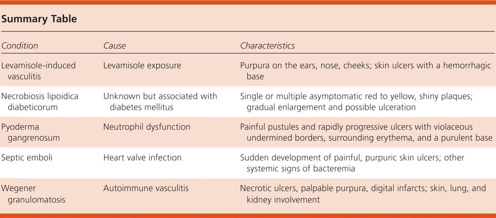
| Condition | Cause | Characteristics |
|---|---|---|
| Levamisole-induced vasculitis | Levamisole exposure | Purpura on the ears, nose, cheeks; skin ulcers with a hemorrhagic base |
| Necrobiosis lipoidica diabeticorum | Unknown but associated with diabetes mellitus | Single or multiple asymptomatic red to yellow, shiny plaques; gradual enlargement and possible ulceration |
| Pyoderma gangrenosum | Neutrophil dysfunction | Painful pustules and rapidly progressive ulcers with violaceous undermined borders, surrounding erythema, and a purulent base |
| Septic emboli | Heart valve infection | Sudden development of painful, purpuric skin ulcers; other systemic signs of bacteremia |
| Wegener granulomatosis | Autoimmune vasculitis | Necrotic ulcers, palpable purpura, digital infarcts; skin, lung, and kidney involvement |
