
A more recent article on mildly elevated liver transaminase levels is available.
Am Fam Physician. 2017;96(11):709-715
Patient information: See related handout on elevated liver enzymes, written by the authors of this article.
Author disclosure: No relevant financial affiliations.
Mild, asymptomatic elevations (less than five times the upper limit of normal) of alanine transaminase and aspartate transaminase levels are common in primary care. It is estimated that approximately 10% of the U.S. population has elevated transaminase levels. An approach based on the prevalence of diseases that cause asymptomatic transaminase elevations can help clinicians efficiently identify common and serious liver disease. The most common causes of elevated transaminase levels are nonalcoholic fatty liver disease and alcoholic liver disease. Uncommon causes include drug-induced liver injury, hepatitis B and C, and hereditary hemochromatosis. Rare causes include alpha1-antitrypsin deficiency, autoimmune hepatitis, and Wilson disease. Extrahepatic sources, such as thyroid disorders, celiac sprue, hemolysis, and muscle disorders, are also associated with mildly elevated transaminase levels. The initial evaluation should include an assessment for metabolic syndrome and insulin resistance (i.e., waist circumference, blood pressure, fasting lipid level, and fasting glucose or A1C level); a complete blood count with platelets; measurement of serum albumin, iron, total iron-binding capacity, and ferritin; and hepatitis C antibody and hepatitis B surface antigen testing. The nonalcoholic fatty liver disease fibrosis score and the alcoholic liver disease/nonalcoholic fatty liver disease index can be helpful in the evaluation of mildly elevated transaminase levels. If testing for common causes is consistent with nonalcoholic fatty liver disease and is otherwise unremarkable, a trial of lifestyle modification is appropriate. If the elevation persists, hepatic ultrasonography and further testing for uncommon causes should be considered.
Mild, asymptomatic elevations of alanine transaminase (ALT) and aspartate transaminase (AST) levels, defined as less than five times the upper limit of normal, are common in primary care. The prevalence of elevated transaminase levels is estimated to be approximately 10%, although less than 5% of these patients have a serious liver disease.1,2 Understanding the epidemiology of each condition that causes asymptomatic elevated transaminase levels can guide the evaluation.3–6 Elevations greater than five times the upper limit of normal should prompt immediate evaluation6 but are beyond the scope of this article.
WHAT IS NEW ON THIS TOPIC: MILDLY ELEVATED LIVER TRANSAMINASE LEVELS
The NAFLD fibrosis score is a calculator that uses clinical data to predict risk of liver-related complications and death from advanced disease. Clinicians should refer patients with a high NAFLD fibrosis score, increased risk of progression, or coexisting chronic liver disease to a gastroenterologist.
In a two-year prospective study in the United Kingdom that included nearly 1,300 primary care patients with abnormal transaminase levels, excluding fatty liver disease (38% of patients), less than 5% of diagnostic workups revealed significant liver disease, and only 17 persons (1.3%) had serious liver disease that required immediate treatment.
NAFLD = nonalcoholic fatty liver disease.
| Clinical recommendation | Evidence rating | References |
|---|---|---|
| Consider gastroenterology referral for patients with persistent elevations of transaminase levels and for those who are at risk of nonalcoholic fatty liver disease progression. | C | 10 |
| Repeat liver enzyme testing is not necessary in the initial workup for elevated transaminase levels. | B | 43 |
| Lifestyle modifications with follow-up are appropriate if history, physical examination, and workup suggest nonalcoholic fatty liver disease. | B | 4–6, 10, 11, 43 |
| If the history and physical examination are unrevealing, clinicians should initiate a stepwise epidemiologic approach to diagnosing the cause of elevated transaminase levels. | C | 3–5 |
Causes of Elevated Liver Transaminase Levels
Hepatocellular damage releases ALT and AST. Elevations in ALT generally are more specific for liver injury, whereas elevations in AST can also be caused by extrahepatic disorders, such as thyroid disorders, celiac sprue, hemolysis, and muscle disorders.7 Normal ALT levels are defined as 29 to 33 IU per L (0.48 to 0.55 μkat per L) for males and 19 to 25 IU per L (0.32 to 0.42 μkat per L) for females.6 The AST:ALT ratio can suggest a specific disease or give insight into liver disease severity. In a study differentiating alcoholic liver disease from nonalcoholic liver disease, alcoholic liver disease was suggested with an AST:ALT ratio greater than 2 (mean AST:ALT values were 152:70; positive likelihood ratio [LR+] = 17, negative likelihood ratio [LR–] = 0.49). On the other hand, nonalcoholic fatty liver disease (NAFLD) was associated with a ratio of less than 1 (mean AST:ALT values were 66:91; LR+ = 80, LR– = 0.2).8 However, causes of mild, asymptomatic elevation of transaminase levels can generally be categorized as common, uncommon, and rare (Table 1).9
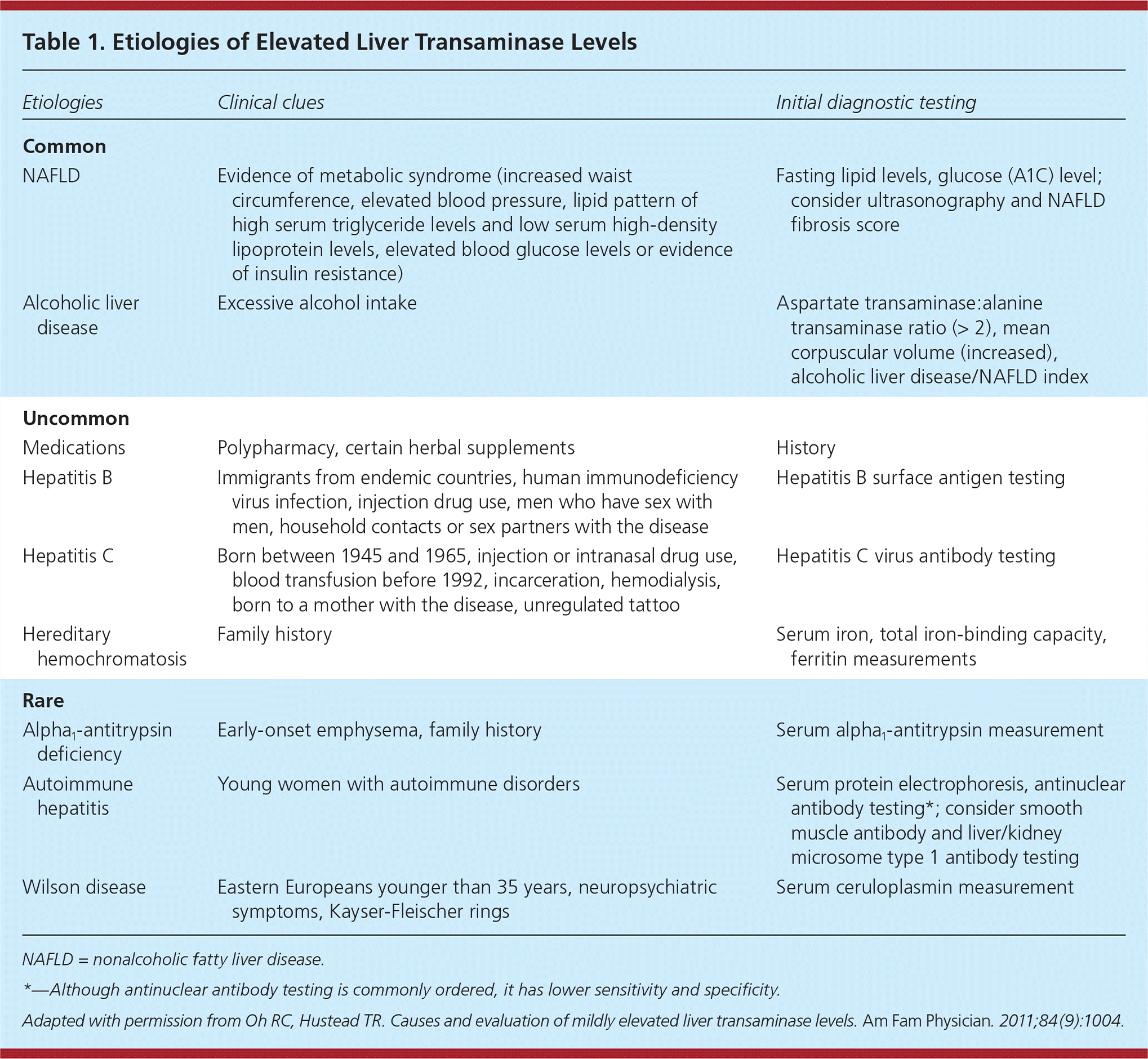
| Etiologies | Clinical clues | Initial diagnostic testing |
|---|---|---|
| Common | ||
| NAFLD | Evidence of metabolic syndrome (increased waist circumference, elevated blood pressure, lipid pattern of high serum triglyceride levels and low serum high-density lipoprotein levels, elevated blood glucose levels or evidence of insulin resistance) | Fasting lipid levels, glucose (A1C) level; consider ultrasonography and NAFLD fibrosis score |
| Alcoholic liver disease | Excessive alcohol intake | Aspartate transaminase:alanine transaminase ratio (> 2), mean corpuscular volume (increased), alcoholic liver disease/NAFLD index |
| Uncommon | ||
| Medications | Polypharmacy, certain herbal supplements | History |
| Hepatitis B | Immigrants from endemic countries, human immunodeficiency virus infection, injection drug use, men who have sex with men, household contacts or sex partners with the disease | Hepatitis B surface antigen testing |
| Hepatitis C | Born between 1945 and 1965, injection or intranasal drug use, blood transfusion before 1992, incarceration, hemodialysis, born to a mother with the disease, unregulated tattoo | Hepatitis C virus antibody testing |
| Hereditary hemochromatosis | Family history | Serum iron, total iron-binding capacity, ferritin measurements |
| Rare | ||
| Alpha1-antitrypsin deficiency | Early-onset emphysema, family history | Serum alpha1-antitrypsin measurement |
| Autoimmune hepatitis | Young women with autoimmune disorders | Serum protein electrophoresis, antinuclear antibody testing*; consider smooth muscle antibody and liver/kidney microsome type 1 antibody testing |
| Wilson disease | Eastern Europeans younger than 35 years, neuropsychiatric symptoms, Kayser-Fleischer rings | Serum ceruloplasmin measurement |
COMMON HEPATIC CAUSES
Nonalcoholic Fatty Liver Disease. A systematic review found that NAFLD is the most common cause of asymptomatic elevation of transaminase levels (25% to 51% of patients with elevated ALT or AST, depending on the study population).1,10 NAFLD is divided into two subtypes. The first is nonalcoholic fatty liver, defined as hepatic steatosis without inflammation. The second, more severe subtype is nonalcoholic steatohepatitis, which is characterized by hepatocyte injury with ballooning of cells, inflammation, and in severe cases, fibrosis.10 Nonalcoholic fatty liver is generally benign and treated successfully with lifestyle modification (Table 211–14), whereas patients with nonalcoholic steatohepatitis have significant risk of progression to cirrhosis and hepatocellular carcinoma. The prevalence of nonalcoholic steatohepatitis is estimated at 3% to 5% of the adult population.10 Thus, the clinical challenge is to determine which patients with NAFLD are at risk of progression.
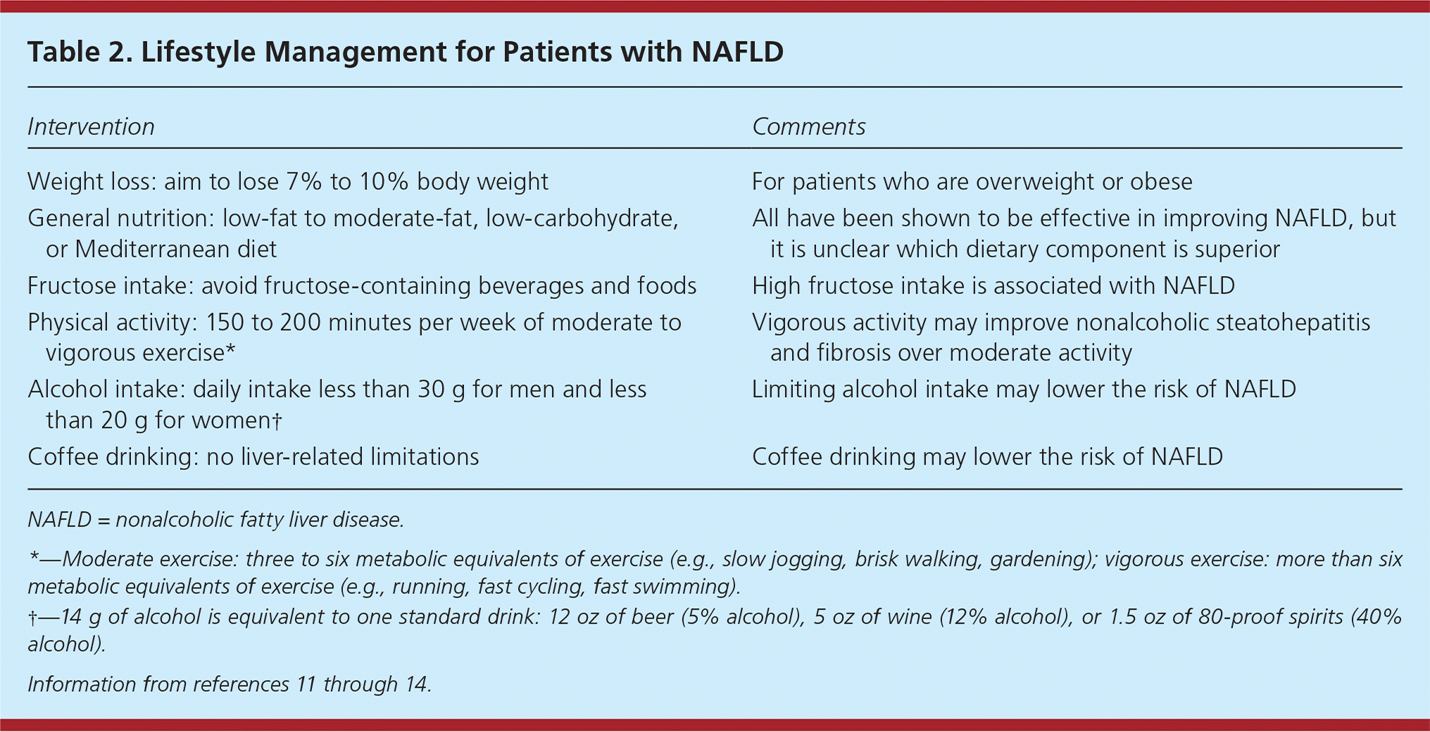
| Intervention | Comments |
|---|---|
| Weight loss: aim to lose 7% to 10% body weight | For patients who are overweight or obese |
| General nutrition: low-fat to moderate-fat, low-carbohydrate, or Mediterranean diet | All have been shown to be effective in improving NAFLD, but it is unclear which dietary component is superior |
| Fructose intake: avoid fructose-containing beverages and foods | High fructose intake is associated with NAFLD |
| Physical activity: 150 to 200 minutes per week of moderate to vigorous exercise* | Vigorous activity may improve nonalcoholic steatohepatitis and fibrosis over moderate activity |
| Alcohol intake: daily intake less than 30 g for men and less than 20 g for women† | Limiting alcohol intake may lower the risk of NAFLD |
| Coffee drinking: no liver-related limitations | Coffee drinking may lower the risk of NAFLD |
Because metabolic syndrome is associated with NAFLD, it should be the leading consideration in individuals with increased waist circumference, elevated blood pressure, high serum triglyceride levels, low serum high-density lipoprotein cholesterol levels, and/ or insulin resistance.11 Type 2 diabetes mellitus is an independent risk factor for NAFLD and increases the risk of nonalcoholic steatohepatitis.15 NAFLD is strongly suggested in a patient with hepatic steatosis on imaging, without significant alcohol history (daily intake less than 30 g for men and less than 20 g for women; 14 g of alcohol is equivalent to one standard drink: 12 oz of beer [5% alcohol], 5 oz of wine [12% alcohol], or 1.5 oz of 80-proof spirits [40% alcohol])16 and no other compelling or coexisting liver disease. Ultrasonography is the preferred first-line imaging modality for diagnosing hepatic steatosis,6,11,17 but it does not readily differentiate between nonalcoholic fatty liver and nonalcoholic steatohepatitis.
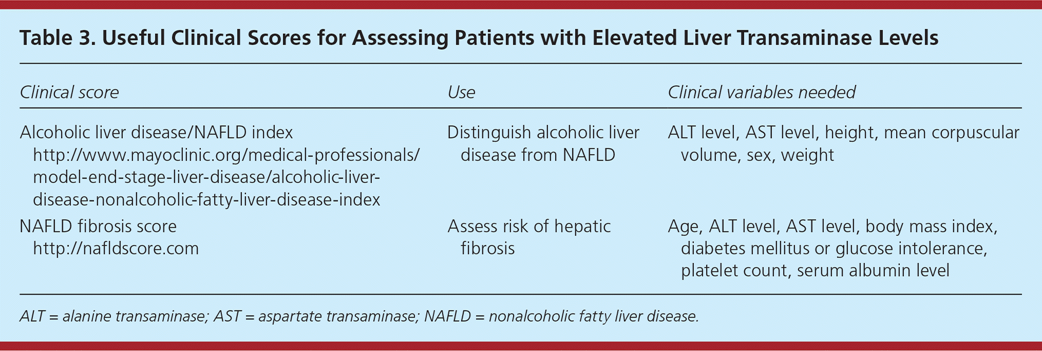
Given the variable progression of NAFLD, it is important to identify those at risk of advanced disease. The presence of liver fibrosis has been shown to be the best predictor of progression.18 Fibrosis is more common in patients older than 50 years.15 A number of clinical tests have been developed to help identify patients with fibrosis in lieu of a liver biopsy. The NAFLD fibrosis score (Table 3) uses clinical data to predict risk of liver-related complications and death from advanced disease.19 Patients with a high NAFLD fibrosis score, increased risk of progression, or coexisting chronic liver disease should be referred to a gastroenterologist.10 Vibration-controlled transient elastography has emerged as a useful noninvasive modality to assess for hepatic fibrosis and may help determine which patients should undergo liver biopsy. However, its use may be limited by operator experience and in patients with elevated body mass index.20
| Clinical score | Use | Clinical variables needed |
|---|---|---|
| Alcoholic liver disease/NAFLD index http://www.mayoclinic.org/medical-professionals/model-end-stage-liver-disease/alcoholic-liver-disease-nonalcoholic-fatty-liver-disease-index | Distinguish alcoholic liver disease from NAFLD | ALT level, AST level, height, mean corpuscular volume, sex, weight |
| NAFLD fibrosis score http://nafldscore.com | Assess risk of hepatic fibrosis | Age, ALT level, AST level, body mass index, diabetes mellitus or glucose intolerance, platelet count, serum albumin level |
Alcoholic Liver Disease. Excessive alcohol intake is the primary cause of liver-related mortality in western countries.21 Alcoholic liver disease and NAFLD have significant overlap in disease spectrum and histopathology.22 If history does not clearly distinguish the two conditions, the alcoholic liver disease/NAFLD index (Table 3) can be used. This index differentiates the conditions based on ALT level, AST level, height, mean corpuscular volume, sex, and weight. It has an LR+ of 12 and an LR– of 0.07, and has been prospectively validated in several varied populations.22–24
UNCOMMON HEPATIC CAUSES
Drug-Induced Liver Injury. The true incidence of drug-induced liver injury is unknown and likely underreported, although it has been estimated at 19.1 cases per 100,000 persons annually.25 A history eliciting prescription and over-the-counter medication use, including supplements, is critical in identifying drug-induced liver injury. With increasing use, supplements now cause 9% of drug-induced liver injury cases.25 Acetaminophen, another common medication, can cause elevated transaminase levels in therapeutic doses.26,27 Medications commonly associated with drug-induced liver injury are listed in Table 4.25,28 The National Institute of Diabetes and Digestive and Kidney Diseases and the National Library of Medicine have collaborated to develop Liver-Tox, a resource for clinical information about drug-induced liver injury.
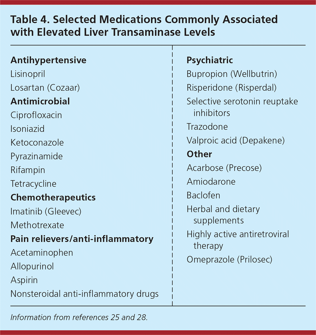
| Antihypertensive |
| Lisinopril |
| Losartan (Cozaar) |
| Antimicrobial |
| Ciprofloxacin |
| Isoniazid |
| Ketoconazole |
| Pyrazinamide |
| Rifampin |
| Tetracycline |
| Chemotherapeutics |
| Imatinib (Gleevec) |
| Methotrexate |
| Pain relievers/anti-inflammatory |
| Acetaminophen |
| Allopurinol |
| Aspirin |
| Nonsteroidal anti-inflammatory drugs |
| Psychiatric |
| Bupropion (Wellbutrin) |
| Risperidone (Risperdal) |
| Selective serotonin reuptake inhibitors |
| Trazodone |
| Valproic acid (Depakene) |
| Other |
| Acarbose (Precose) |
| Amiodarone |
| Baclofen |
| Herbal and dietary supplements |
| Highly active antiretroviral therapy |
| Omeprazole (Prilosec) |
Statin-induced liver injuries are rare.29 Given the lack of evidence linking statins to elevated transaminase levels, the U.S. Food and Drug Administration now recommends only baseline measurement of ALT and AST before initiation of statins, and does not recommend routine liver monitoring for patients taking statins.29 However, clinicians should consider testing if there is suspicion for drug-induced liver injury or other liver disease. Statins have also been shown to be safe in stable chronic liver disease such as NAFLD and hepatitis C.25
Viral Hepatitis. Hepatitis B and C are common causes of elevated transaminase levels.1 In the United States, approximately 3.5 million persons have chronic hepatitis C virus infection, and up to 2.2 million have hepatitis B virus infection.30 The U.S. Preventive Services Task Force recommends screening high-risk patients with hepatitis B surface antigen and hepatitis C virus antibody testing.31,32
Hereditary Hemochromatosis. Hereditary hemochromatosis is an autosomal recessive disease causing increased iron absorption in the intestines and release by tissue macrophages. Although the classic gene mutation is common in Northern European Caucasians, at one per 150 to 250 persons, the severe iron overload of hereditary hemochromatosis is only phenotypically expressed in approximately 10% of patients with the genotype.33,34
Mild, asymptomatic elevations in liver enzymes can occur because iron itself does not elicit a significant inflammatory response in the liver. Transferrin saturation and serum ferritin level should be measured to rule out hereditary hemochromatosis in patients with elevated transaminase levels. Transferrin saturation of 45% or more, and serum ferritin levels of more than 250 to 300 ng per mL (562 to 674 pmol per Lng per mL) in men or more than 200 ng per mL (449 pmol per Lng per mL) in women should prompt testing for the presence of the HFE gene.33 Higher thresholds for transferrin saturation (more than 60% in men and more than 50% in women) have been shown to predict the presence of hereditary hemochromatosis with 95% accuracy.35 The homozygous C282Y gene mutation is responsible for 80% to 85% of cases.33
RARE CAUSES
Alpha1-Antitrypsin Deficiency. Alpha1-antitrypsin deficiency is a genetic condition that primarily causes chronic lung and liver disease. The prevalence is approximately one in 3,000 to 5,000 persons, but only 10% of those with the disease are diagnosed.36 In the liver, an accumulation of an abnormal alpha1-antitrypsin protein results in progressive damage. Although there are more than 100 alpha1-antitrypsin gene variants, more than 95% of clinical disease is in ZZ homozygotes, also known as the PiZZ genotype.37,38 Alpha1-antitrypsin deficiency should be suspected in patients with early-onset emphysema, and elevations in liver enzymes or clinical findings of advanced liver disease without a known cause. Diagnosis begins with testing for serum alpha1-antitrypsin deficiency. If levels are very low, protein phenotyping or genotyping to look for the PiZZ variant should follow.36
Autoimmune Hepatitis. The prevalence of autoimmune hepatitis is 11 to 17 per 100,000 persons.39 It occurs more often in young women and is associated with other autoimmune disorders. Hypergammaglobulinemia is common in patients with autoimmune hepatitis, with total immunoglobulin G levels generally 1.2 to 3 times normal.40 Therefore, serum protein electrophoresis testing has high sensitivity for autoimmune hepatitis, ruling out the condition if results are normal. Although antinuclear antibody testing is commonly ordered, it has lower sensitivity and specificity.41 Other laboratory tests may include smooth muscle antibody and liver/kidney microsome type 1 antibody measurements.39
Wilson Disease. Wilson disease is a rare autosomal recessive disorder, occurring in approximately one in 30,000 persons, and is related to ineffective copper metabolism. It usually occurs in Eastern Europeans younger than 35 years.42 Kayser-Fleischer rings (copper deposition around the cornea) or neuropsychiatric symptoms are key clinical clues. A serum ceruloplasmin measurement is the initial test.42 If ceruloplasmin levels are low, further investigation with 24-hour urine copper levels, genetic testing, and liver biopsy can be considered.
Extrahepatic Causes. A number of extrahepatic sources of asymptomatic transaminase elevations may be considered based on the clinical picture.6 For example, thyroid disorders and celiac sprue have been associated with elevated transaminase levels.4 If the clinical picture is consistent, hemolysis and strenuous exercise should be considered.7 Rhabdomyolysis and polymyositis are unlikely etiologies, but creatine kinase or aldolase measurements should be considered in patients with significant myalgias.
Suggested Diagnostic Evaluation
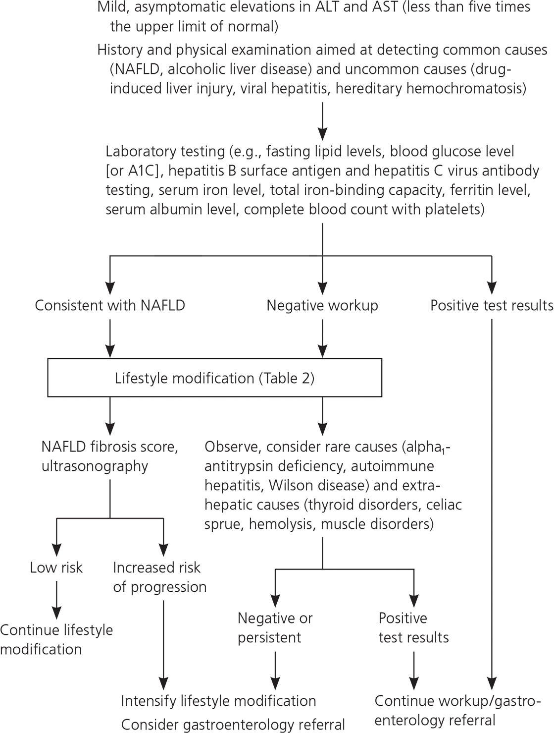
A large prospective study performed in the United Kingdom evaluated nearly 1,300 primary care patients with abnormal transaminase levels or liver function testing for two years to determine the cause of the abnormalities.43 Each patient underwent laboratory testing and liver ultrasonography. Excluding fatty liver disease (found in 38% of the cohort), less than 5% of diagnostic workups revealed significant liver disease. Only 17 (1.3%) of 1,300 patients had serious liver disease that required immediate treatment—13 cases of viral hepatitis (1%) and four cases of hereditary hemochromatosis (0.3%). Notably, repeating liver enzyme testing did not appear to be efficient because 84% of results remained abnormal after one month and 75% after two years. This study, along with guidelines, informs the evaluation of mildly elevated transaminase levels in primary care (Figure 1).3–6,43
Data Sources: A PubMed search was completed using the key terms elevated, liver function tests, transaminases, and aminotransferases. In addition, we used the key words nonalcoholic fatty liver disease, alcoholic liver disease, viral hepatitis, hemochromatosis, alpha1-antitrypsin deficiency, autoimmune hepatitis, and Wilson's disease alone or in combination with aminotransferases. The search included the “diagnosis” clinical study category and related articles in PubMed. Also searched were Essential Evidence and the updated guidelines from the American Association for the Study of Liver Disease. Search dates: May 1, 2016, to April 8, 2017.
The views expressed in this manuscript are those of the authors and do not reflect the official policy or position of the Department of the Army, the Department of Defense, or the U.S. government.
