
Am Fam Physician. 2018;98(7):421-428
Patient information: See related handout on low back pain.
Author disclosure: No relevant financial affiliations.
Low back pain is usually nonspecific or mechanical. Mechanical low back pain arises intrinsically from the spine, intervertebral disks, or surrounding soft tissues. Clinical clues, or red flags, may help identify cases of nonmechanical low back pain and prompt further evaluation or imaging. Red flags include progressive motor or sensory loss, new urinary retention or overflow incontinence, history of cancer, recent invasive spinal procedure, and significant trauma relative to age. Imaging on initial presentation should be reserved for when there is suspicion for cauda equina syndrome, malignancy, fracture, or infection. Plain radiography of the lumbar spine is appropriate to assess for fracture and bony abnormality, whereas magnetic resonance imaging is better for identifying the source of neurologic or soft tissue abnormalities. There are multiple treatment modalities for mechanical low back pain, but strong evidence of benefit is often lacking. Moderate evidence supports the use of nonsteroidal anti-inflammatory drugs, opioids, and topiramate in the short-term treatment of mechanical low back pain. There is little or no evidence of benefit for acetaminophen, antidepressants (except duloxetine), skeletal muscle relaxants, lidocaine patches, and transcutaneous electrical nerve stimulation in the treatment of chronic low back pain. There is strong evidence for short-term effectiveness and moderate-quality evidence for long-term effectiveness of yoga in the treatment of chronic low back pain. Various spinal manipulative techniques (osteopathic manipulative treatment, spinal manipulative therapy) have shown mixed benefits in the acute and chronic setting. Physical therapy modalities such as the McKenzie method may decrease the recurrence of low back pain and use of health care. Educating patients on prognosis and incorporating psychosocial components of care such as identifying comorbid psychological problems and barriers to treatment are essential components of long-term management.
Mechanical low back pain refers to back pain that arises intrinsically from the spine, intervertebral disks, or surrounding soft tissues. This includes lumbosacral muscle strain, disk herniation, lumbar spondylosis, spondylolisthesis, spondylolysis, vertebral compression fractures, and acute or chronic traumatic injury.1 Repetitive trauma and overuse are common causes of chronic mechanical low back pain, which is often secondary to workplace injury. Most patients who experience activity-limiting low back pain go on to have recurrent episodes. Chronic low back pain affects up to 23% of the population worldwide, with an estimated 24% to 80% of patients having a recurrence at one year.2,3
| Clinical recommendation | Evidence rating | References |
|---|---|---|
| Do not order initial imaging studies unless there is concern for cauda equina syndrome, malignancy, fracture, or infection. | B | 5, 6, 8–11 |
| Nonsteroidal anti-inflammatory drugs, opioids, and topiramate (Topamax) are more effective than placebo in the short-term treatment of nonspecific chronic low back pain. | A | 12, 13, 15, 16 |
| Acetaminophen, antidepressants (except duloxetine [Cymbalta]), lidocaine patches, and transcutaneous electrical nerve stimulation are not consistently more effective than placebo in the treatment of chronic low back pain. | B | 12, 18, 20, 22, 33 |
| Consider referral to physical therapy for McKenzie method techniques to reduce the risk of recurrence and need for health care services. | B | 24, 25, 45–47 |
| Intensive patient education that includes advice to stay active, avoid aggravating movements, and return to normal activity as soon as possible, and a discussion of the often benign nature of acute low back pain is effective in patients with nonspecific pain. | B | 36 |
| Recommendation | Sponsoring organization |
|---|---|
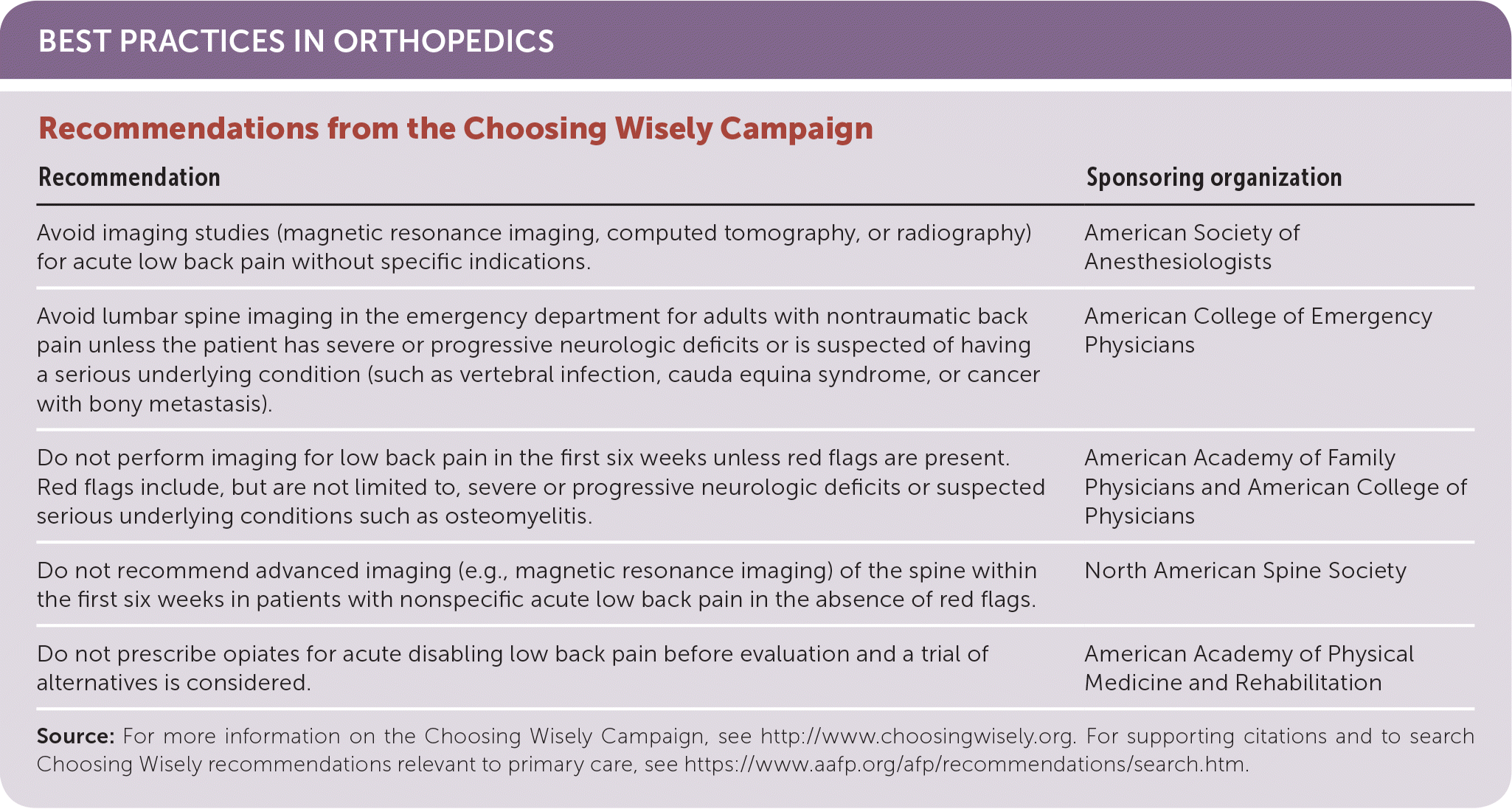
| Avoid imaging studies (magnetic resonance imaging, computed tomography, or radiography) for acute low back pain without specific indications. | American Society of Anesthesiologists |
| Avoid lumbar spine imaging in the emergency department for adults with nontraumatic back pain unless the patient has severe or progressive neurologic deficits or is suspected of having a serious underlying condition (such as vertebral infection, cauda equina syndrome, or cancer with bony metastasis). | American College of Emergency Physicians |
| Do not perform imaging for low back pain in the first six weeks unless red flags are present. Red flags include, but are not limited to, severe or progressive neurologic deficits or suspected serious underlying conditions such as osteomyelitis. | American Academy of Family Physicians and American College of Physicians |
| Do not recommend advanced imaging (e.g., magnetic resonance imaging) of the spine within the first six weeks in patients with nonspecific acute low back pain in the absence of red flags. | North American Spine Society |
| Do not prescribe opiates for acute disabling low back pain before evaluation and a trial of alternatives is considered. | American Academy of Physical Medicine and Rehabilitation |
Medicare expenditures for patients with low back pain have increased dramatically, with large increases in spending on epidural corticosteroid injections and opiate prescriptions (629% and 423% increases, respectively), as well as increased use of magnetic resonance imaging and spinal fusion surgery, without any significant improvement in patient outcomes or disability rates.4 This article proposes a more judicious use of imaging, with use of evidence-based treatments to decrease cost, decrease disability, and improve overall outcomes.
Evaluation
The history and physical examination, with appropriate use of imaging, can point toward a specific etiology. However, the complexity and biomechanics of the spine make it difficult to identify a specific anatomic lesion, with a precise diagnosis made in only 20% of cases.5
HISTORY AND PHYSICAL EXAMINATION
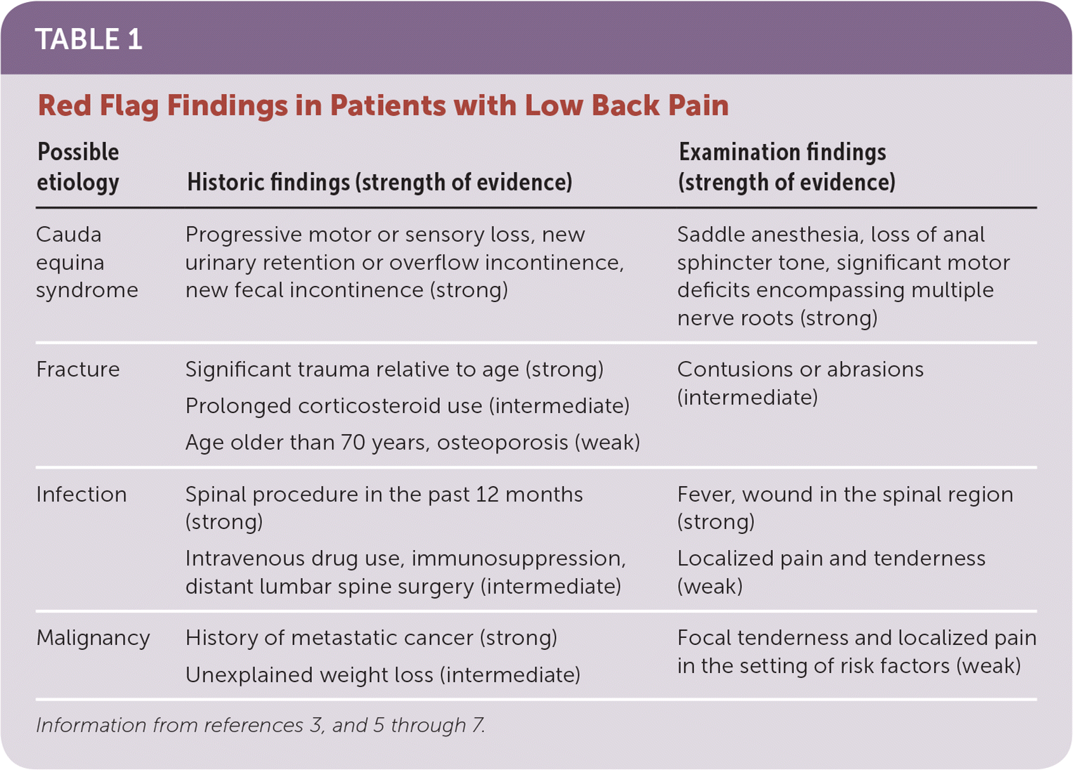
Evaluation of low back pain should begin with a history and physical examination, the results of which dictate further evaluation or treatment. The presence of red flags that suggest systemic disease or urgent problems warrants additional evaluation before empiric treatment (Table 13,5–7). A systematic review showed a higher likelihood of fracture with the presence of one or more red flags for trauma (older age, prolonged corticosteroid use, significant trauma relative to age, contusions or abrasions). History of malignancy had the highest posttest probability for detection of spinal malignancy.6 Other important red flags include constitutional symptoms for malignancy or infection, loss of bowel or bladder function and progressive motor or sensory loss for cauda equina syndrome, and history of a spinal procedure or intravenous drug use for infection.
| Possible etiology | Historic findings (strength of evidence) | Examination findings (strength of evidence) |
|---|---|---|
| Cauda equina syndrome | Progressive motor or sensory loss, new urinary retention or overflow incontinence, new fecal incontinence (strong) | Saddle anesthesia, loss of anal sphincter tone, significant motor deficits encompassing multiple nerve roots (strong) |
| Fracture | Significant trauma relative to age (strong) Prolonged corticosteroid use (intermediate) | Contusions or abrasions (intermediate) |
| Age older than 70 years, osteoporosis (weak) | ||
| Infection | Spinal procedure in the past 12 months (strong) | Fever, wound in the spinal region (strong) |
| Intravenous drug use, immunosuppression, distant lumbar spine surgery (intermediate) | Localized pain and tenderness (weak) | |
| Malignancy | History of metastatic cancer (strong) | Focal tenderness and localized pain in the setting of risk factors (weak) |
| Unexplained weight loss (intermediate) |
The history should include an assessment of pain location, severity, timing, aggravating/relieving factors, and radiation. Body mass index, physical activity, and occupational hazards should be used to assess risk of mechanical low back pain. Patients with psychosocial deficits or disabilities are more likely to develop chronic back pain and are more likely to be disabled by their symptoms.1
Physical examination should include evaluation of strength, sensation, and reflexes of the lower extremities. Inspection, palpation, and range-of-motion testing of the lumbosacral musculature are helpful for identifying point tenderness, restriction, and spasm. The straight leg raise test is performed by the examiner raising the patient's straight leg to an angle of 30 to 70 degrees. Ipsilateral leg pain at less than 60 degrees is positive for lumbar disk herniation (sensitivity = 0.80, specificity = 0.40; positive likelihood ratio = 2.0, negative likelihood ratio = 0.5). Reproduction of contralateral pain using the crossed straight leg raise test is positive for lumbar disk herniation (sensitivity = 0.35, specificity = 0.90; positive likelihood ratio = 3.5, negative likelihood ratio = 0.72).7 Patients with psychosocial symptoms or risk factors may be assessed for nonorganic or inappropriate physical signs.
IMAGING
Imaging is not recommended for most patients with nonspecific mechanical low back pain in the absence of red flags.5,6 The American College of Radiology Appropriateness Criteria for low back pain recommends imaging only if there is no improvement after six weeks of conservative medical and physical therapies, or there is high suspicion for cauda equina syndrome, malignancy, fracture, or infection.8 The presence of radiculopathy with low back pain is not an indication for early imaging.8,9 Early imaging with plain radiography is associated with worse overall outcomes and is likely to identify minor abnormalities in asymptomatic patients.8 Plain radiography of the lumbar spine is appropriate to assess for fracture and bony abnormality, whereas magnetic resonance imaging is better for identifying the source of neurologic or soft tissue abnormalities. A randomized trial showed that early routine use of magnetic resonance imaging increases lumbar spinal surgeries without reciprocal benefits in pain or function.10 It is appropriate to begin symptomatic therapy without imaging in adults younger than 50 years and in those older than 50 years if there is not concern for systemic disease.11
DIFFERENTIAL DIAGNOSIS
| Diagnosis (percentage of patients with low back pain) | Key characteristics and clinical clues |
|---|---|
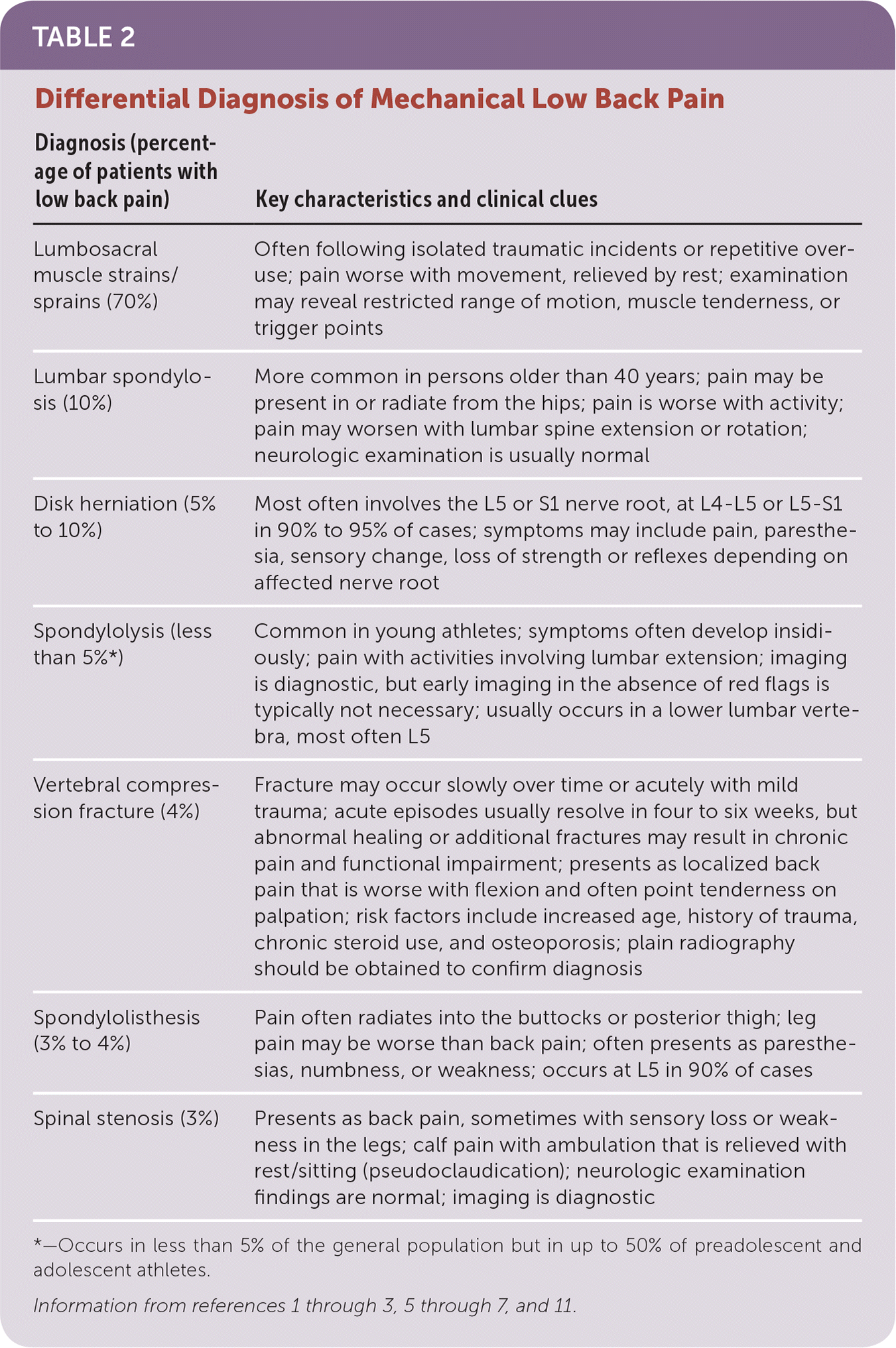
| Lumbosacral muscle strains/sprains (70%) | Often following isolated traumatic incidents or repetitive overuse; pain worse with movement, relieved by rest; examination may reveal restricted range of motion, muscle tenderness, or trigger points |
| Lumbar spondylosis (10%) | More common in persons older than 40 years; pain may be present in or radiate from the hips; pain is worse with activity; pain may worsen with lumbar spine extension or rotation; neurologic examination is usually normal |
| Disk herniation (5% to 10%) | Most often involves the L5 or S1 nerve root, at L4–L5 or L5–S1 in 90% to 95% of cases; symptoms may include pain, paresthesia, sensory change, loss of strength or reflexes depending on affected nerve root |
| Spondylolysis (less than 5%*) | Common in young athletes; symptoms often develop insidiously; pain with activities involving lumbar extension; imaging is diagnostic, but early imaging in the absence of red flags is typically not necessary; usually occurs in a lower lumbar vertebra, most often L5 |
| Vertebral compression fracture (4%) | Fracture may occur slowly over time or acutely with mild trauma; acute episodes usually resolve in four to six weeks, but abnormal healing or additional fractures may result in chronic pain and functional impairment; presents as localized back pain that is worse with flexion and often point tenderness on palpation; risk factors include increased age, history of trauma, chronic steroid use, and osteoporosis; plain radiography should be obtained to confirm diagnosis |
| Spondylolisthesis (3% to 4%) | Pain often radiates into the buttocks or posterior thigh; leg pain may be worse than back pain; often presents as paresthesias, numbness, or weakness; occurs at L5 in 90% of cases |
| Spinal stenosis (3%) | Presents as back pain, sometimes with sensory loss or weakness in the legs; calf pain with ambulation that is relieved with rest/sitting (pseudoclaudication); neurologic examination findings are normal; imaging is diagnostic |
Treatment
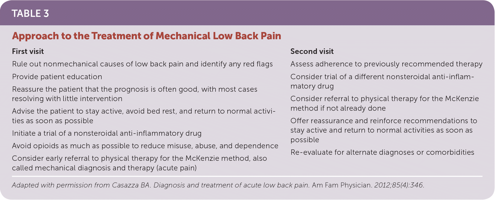
Multiple treatments may be employed for acute or chronic mechanical low back pain, although strong evidence for their benefit is lacking.5 Once nonmechanical causes of low back pain are ruled out, a stepwise approach to management is recommended (Table 35). Treatment options are summarized in Table 4.12–36
| First visit | Second visit |
|---|---|
| Rule out nonmechanical causes of low back pain and identify any red flags Provide patient education Reassure the patient that the prognosis is often good, with most cases resolving with little intervention Advise the patient to stay active, avoid bed rest, and return to normal activities as soon as possible Initiate a trial of a nonsteroidal anti-inflammatory drug Avoid opioids as much as possible to reduce misuse, abuse, and dependence Consider early referral to physical therapy for the McKenzie method, also called mechanical diagnosis and therapy (acute pain) | Assess adherence to previously recommended therapy Consider trial of a different nonsteroidal anti-inflammatory drug Consider referral to physical therapy for the McKenzie method if not already done Offer reassurance and reinforce recommendations to stay active and return to normal activities as soon as possible Re-evaluate for alternate diagnoses or comorbidities |

| Pharmacologic | |
| Acetaminophen | No evidence that acetaminophen is better than placebo 12 |
| Opioid-sparing or synergistic effects may justify use despite lack of high-quality evidence | |
| Nonsteroidal anti-inflammatory drugs | Effective for short-term relief of chronic low back pain without radiculopathy12 |
| No difference between NSAIDs and placebo for radicular symptoms13 | |
| No difference between different types of NSAIDs and between NSAIDs and other commonly used pharmacotherapies, including opioids and muscle relaxants, when used for chronic pain13 | |
| Anticonvulsants | Gabapentinoids have significant adverse effects without demonstrated benefits in patients with chronic low back pain14 |
| A single trial with 96 patients concluded that topiramate (Topamax) was more effective than placebo in improving pain severity or functioning in patients with chronic low back pain15 | |
| Opioids | Short-term effectiveness for pain relief and functioning, but long-term effectiveness and safety are unclear |
| Increased risk of misuse, abuse, and diversion16,17 | |
| Skeletal muscle relaxants | More effective pain relief and global efficacy in acute chronic nonspecific low back pain when compared with placebo; however, adverse effects such as sedation, abuse (carisoprodol [Soma]), transiently lower blood pressure (tizanidine [Zanaflex]), and increased risk of serotonin syndrome (cyclobenzaprine [Flexeril]) are common18 |
| No additional benefit when added to naproxen19 | |
| Topical anesthetics | Topical lidocaine patches appear to be no more effective than placebo20 |
| Oral corticosteroids | No benefit for acute low back pain according to a single randomized controlled trial21 |
| Antidepressants | No clear evidence of superiority over placebo for chronic low back pain to support the use of antidepressants, except for duloxetine (Cymbalta), in patients with comorbid depression or other forms of chronic pain22 |
| Physical | |
| McKenzie method (mechanical diagnosis and therapy) | Initial assessment by a physical therapist trained in the methodology followed by an individualized self-treatment program has been shown to have moderate evidence for acute low back pain but moderate to no difference for chronic low back pain23–25 |
| Osteopathic manipulative treatment | Shown to be effective at reducing acute and chronic mechanical low back pain in a systematic review and meta-analysis26 |
| Acupuncture and dry needling | Small benefit when added to conventional therapies27 |
| Massage | Low- to very-low-quality evidence that massage may lead to short-term improvements in pain outcomes for acute, subacute, and chronic low back pain28 |
| Surgical/invasive | |
| Surgery | Consider referral for surgery in patients who have had disabling low back pain impacting quality of life for more than one year29 |
| Injection therapy | Insufficient evidence to support injection therapy (i.e., corticosteroids, anesthetics, and other drugs administered at epidural sites, facet joints, or local sites) in subacute and chronic low back pain30 |
| Prolotherapy injections alone are not effective for chronic low back pain but may improve chronic pain when used in conjunction with other treatments31 | |
| Psychological and other modalities | |
| Cognitive behavior therapy | Greater improvement in back pain and functional limitations if incorporated into treatment plans for chronic low back pain32 |
| Transcutaneous electrical nerve stimulation | No functional benefit in the treatment of patients with chronic low back pain33 |
| Yoga | Strong evidence of short-term effectiveness and moderate-quality evidence of long-term effectiveness for chronic low back pain34 |
| Multidisciplinary rehabilitation | More effective than usual care35 |
| Patient education | Strong evidence that intensive, 2.5-hour educational sessions (e.g., advice to stay active, avoid aggravating movements, and return to normal activity as soon as possible, and a discussion of the often benign nature of acute low back pain) are more effective for return to work and long-term pain in patients with acute or subacute low back pain36 |
| Less intensive patient education is no more effective than no intervention, and comparison of different types of education did not show significant differences36 | |
PHARMACOLOGIC
Nonsteroidal anti-inflammatory drugs (NSAIDs) are often used as first-line treatment and may provide short-term relief. NSAIDs are effective for short-term relief in chronic low back pain without radiculopathy, but there is no difference between NSAIDs and placebo for radicular symptoms.12 There is also no difference in effectiveness between different types of NSAIDs and between NSAIDs and other commonly used pharmacotherapies, including opioids and muscle relaxants, in those with chronic pain.13,16 There is no evidence that acetaminophen is better than placebo.12 Moderate-quality evidence suggests that skeletal muscle relaxants are beneficial in nonspecific chronic low back pain. However, adverse effects, including sedation, abuse, transient hypotension, and serotonin syndrome, may limit their use.18 Two recent randomized controlled trials did not show a benefit from systemic corticosteroids compared with placebo in patients with acute low back pain caused by a herniated lumbar disk37 or a benefit from cyclobenzaprine (Flexeril) plus an NSAID compared with an NSAID alone in patients with nonradicular acute low back pain.19
PHYSICAL THERAPY AND MANUAL MEDICINE
Physical therapists play an integral role in the diagnosis and treatment of low back pain; variable evidence exists for specific physical modalities. Manipulation and mobilization are no more effective than inert interventions for acute low back pain.38 However, a systematic review and meta-analysis concluded that osteopathic manipulative treatment is effective in reducing acute and chronic mechanical low back pain.26 There is strong evidence that spinal stabilization exercises have no long-term advantages over other exercises.39 Exercise therapy is as effective as other therapies for the treatment of acute low back pain, and is slightly effective at reducing pain and improving function in chronic low back pain.40 However, early guideline-directed physical therapy has substantial reductions in use of health care and overall costs.41
SURGICAL/INVASIVE
A Cochrane review concluded that there is no strong evidence for or against the use of injection therapy (i.e., corticosteroids, anesthetics, and other drugs administered at epidural sites, facet joints, or local sites) in the treatment of low back pain.30 Relatively few patients with mechanical low back pain will benefit from surgery. Other than indications for urgent surgical referral, such as progressive motor weakness or cauda equina syndrome, the American Pain Society recommends offering surgery only to patients who have had disabling low back pain impacting quality of life for more than one year.29 Spinal fusion and lumbar disk replacement are the most common procedures for mechanical low back pain. However, evidence of superiority over nonsurgical modalities is limited,29,42 and a randomized trial found no clear benefit of spinal fusion after nearly 13 years of follow-up.43
PSYCHOLOGICAL AND OTHER MODALITIES
Cognitive behavior therapy and mindfulness-based stress reduction may play a role in the treatment of chronic low back pain, with improvements in back pain and functional limitations after 26 weeks of treatment.32 There is strong evidence for short-term effectiveness and moderate-quality evidence for long-term effectiveness of yoga in the treatment of chronic low back pain.34 Engaging in multidisciplinary biopsychosocial rehabilitation can result in decreased pain and disability secondary to low back pain, with moderate-quality evidence suggesting that it is more effective than usual care.35
MCKENZIE METHOD
The McKenzie method (mechanical diagnosis and therapy) is a physical therapy approach that uses a structured examination to classify patients with low back pain, which helps identify those who will benefit from physical therapy and which treatment will provide the most benefit.44 Classification is based on an assessment of the patient's symptomatic and mechanical responses from repeated or sustained end-range-of-motion joint movements.44 For example, a patient might report decreased radicular symptoms after performing repeated standing lumbar extension movements.
The McKenzie method describes two key examination findings: the centralization phenomenon and directional preference. The centralization phenomenon is rapid change in the location of pain from a peripheral or distal location to a more proximal or central location in response to treatment (e.g., a patient describing pain in the foot that moves out of the foot and into the buttock with repeated movements).45 Patients who demonstrate centralization at initial evaluation have better outcomes, which can be used as a prognostic indicator.46 Directional preference is a rapid and lasting positive change in symptom, function, or range of motion occurring from repeated end range joint movement testing in one specific direction of movement. Directional preference describes the direction of spinal movement or position that produces centralization; however, it is not synonymous with centralization.45 Directional preference is determined from a decrease in pain intensity without a change in pain location. For example, a patient who performs repeated lumbar extensions while standing reports decreased leg symptom intensity. This identifies a directional preference for extension even without centralization.
A randomized controlled trial of 241 patients found that when patients with directional preference or centralization are matched with a treatment in the same direction, they are more likely to have a positive outcome, defined as at least a 30% decrease on the Roland Morris Disability Questionnaire (odds ratio of 7.8 vs. 0.77 in the unmatched group).45 Those with directional preference or centralization who are prescribed unmatched exercises (exercises in the opposite direction) have negative outcomes. This supports the idea that matched diagnosis and treatment are key to positive outcomes.
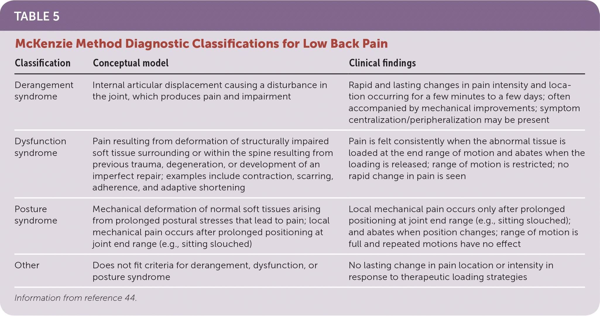
The McKenzie method has four main diagnostic classifications: derangement syndrome, dysfunction syndrome, posture syndrome, and other.44 Clinicians trained in the McKenzie method evaluate patients based on history findings, establishing mechanical baselines and performing an active movement examination to evaluate responses as described earlier. Each classification has different hallmark features, and each requires different treatment approaches.23 After patients are classified, they are taught specific, individualized, and primarily self-treatment exercises to perform at home, which promotes independence and patient ownership. Each patient is given a prescription for frequency and duration of exercises based on his or her classification, including expectations, safety guidelines, and warning signs. Table 5 explains the conceptual model and clinical findings relevant to each classification.44
| Classification | Conceptual model | Clinical findings |
|---|---|---|
| Derangement syndrome | Internal articular displacement causing a disturbance in the joint, which produces pain and impairment | Rapid and lasting changes in pain intensity and location occurring for a few minutes to a few days; often accompanied by mechanical improvements; symptom centralization/peripheralization may be present |
| Dysfunction syndrome | Pain resulting from deformation of structurally impaired soft tissue surrounding or within the spine resulting from previous trauma, degeneration, or development of an imperfect repair; examples include contraction, scarring, adherence, and adaptive shortening | Pain is felt consistently when the abnormal tissue is loaded at the end range of motion and abates when the loading is released; range of motion is restricted; no rapid change in pain is seen |
| Posture syndrome | Mechanical deformation of normal soft tissues arising from prolonged postural stresses that lead to pain; local mechanical pain occurs after prolonged positioning at joint end range (e.g., sitting slouched) | Local mechanical pain occurs only after prolonged positioning at joint end range (e.g., sitting slouched); and abates when position changes; range of motion is full and repeated motions have no effect |
| Other | Does not fit criteria for derangement, dysfunction, or posture syndrome | No lasting change in pain location or intensity in response to therapeutic loading strategies |
The McKenzie method has moderate evidence of effectiveness in reducing pain and improving function in patients with low back pain.23–25 Reliability is increased with training and experience with the classification system. Clinicians trained in the McKenzie method demonstrate good inter-rater reliability when classifying patients.47,48 Physicians, physical therapists, nurse practitioners, physician assistants, and chiropractors are eligible for training. In case reports, physical therapists with training have been clinically proven to demonstrate good pain and function outcomes for patients with low back pain.23 Training in the McKenzie method consists of two levels of certification and multiple components. Information on education, credentialing, and locating trained clinicians, go to http://mckenzieinstituteusa.org.
Data Sources: We searched PubMed for the key term low back pain, with mechanical, McKenzie, MDT, physical therapy, red flags, differential diagnosis, manipulation, treatment, NSAIDs, muscle relaxants, steroids, acetaminophen, exercise, disability. We also searched the Cochrane Database of Systematic Reviews, Clinical Evidence, Essential Evidence Plus, and the National Guideline Clearinghouse. Search dates: March to May 2017, and May 2018.
The opinions and assertions contained herein are the private views of the authors and are not to be construed as official or as reflecting the views of the U.S. Army Medical Department or the U.S. Army Service at large.
