Am Fam Physician. 2019;99(5):327-328
Author disclosure: No relevant financial affiliations.
A 29-year-old man presented with a painless, white area on the roof of his mouth that had been present for three months. He had no difficulty with eating or drinking. He was otherwise healthy with no medical conditions, although he had poor dentition. He had smoked cigarettes and used chewing tobacco for 11 years.
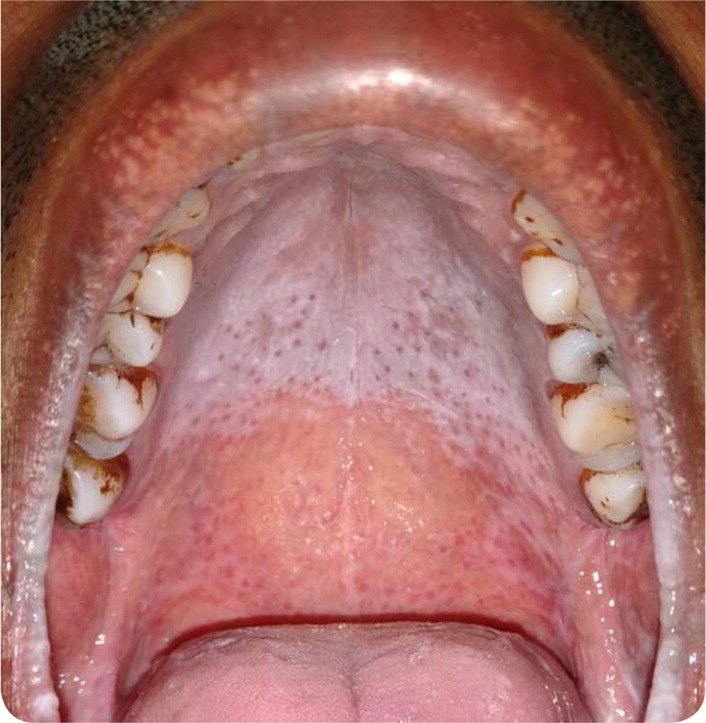
The oral examination revealed multiple brown to red punctate lesions against a white background on the palate (Figure 1).

Question
Discussion
The answer is B: nicotine stomatitis. Tobacco and its combustion products can cause a wide variety of lesions in the oral cavity, including leukoplakia, leukoedema, black hairy tongue, and nicotine stomatitis. Nicotine stomatitis commonly occurs in pipe smokers because the heat from the pipe stem is directed toward the palate. It can also occur in cigarette smokers who reverse smoke and in people who drink hot tea and coffee. The classic presentation is multiple red, umbilicated papules against a gray-white background (smoker's palate).1
The brown to red punctate lesions are inflamed mucous gland orifices, which predominate over the posterior part of the hard palate. The diffuse white background is secondary to increased keratinization. Histopathology shows hyperkeratosis, parakeratosis, and acanthosis1 and may demonstrate a chronic sialodochitis (inflammation of the salivary ducts). Nicotine stomatitis lesions are asymptomatic and usually resolve with cessation of smoking.
Leukoedema presents as asymptomatic white or whitish-gray, translucent reticulations on the buccal and labial mucosa. It is commonly associated with a local irritation. The lesions disappear when the mucosa is stretched.2
Oral candidiasis, or thrush, commonly presents as curdy, yellowish-white plaques. It is caused by a yeast or fungal infection of the mucous membranes in the mouth. Candida albicans is the most common causative organism. A potassium hydroxide preparation from lesion scrapings is useful in identifying the infection.
Oral lichen planus is an inflammatory condition. The reticular form, the most common variant of oral lichen planus, presents as white lacy patches and plaques on the buccal mucosa and the tongue.3 Skin and other mucosal sites may also be involved.

| Condition | Characteristics |
|---|---|
| Leukoedema | Asymptomatic white or whitish-gray, translucent reticulations on the buccal and labial mucosa; the lesions disappear when the mucosa is stretched |
| Nicotine stomatitis | Heat-related injury to the palate common in pipe and cigarette smokers and people who drink hot beverages; brown to red punctate lesions on a gray-white background |
| Oral candidiasis | Yeast or fungal infection most commonly caused by Candida albicans; curdy, yellowish-white plaques; a potassium hydroxide preparation from lesion scrapings is useful in identifying the infection |
| Oral lichen planus | Inflammatory condition; the reticular variant (most common) presents as white lacy patches and plaques on the buccal mucosa and tongue; skin and other mucosal sites may also be involved |
