Am Fam Physician. 2019;99(8):490-496
Author disclosure: No relevant financial affiliations.
Kidney stones are a common disorder, with an annual incidence of eight cases per 1,000 adults. During an episode of renal colic, the first priority is to rule out conditions requiring immediate referral to an emergency department, then to alleviate pain, preferably with a nonsteroidal anti-inflammatory drug. The diagnostic workup consists of urinalysis, urine culture, and imaging to confirm the diagnosis and assess for conditions requiring active stone removal, such as urinary infection or a stone larger than 10 mm. Conservative management consists of pain control, medical expulsive therapy with an alpha blocker, and follow-up imaging within 14 days to monitor stone position and assess for hydronephrosis. Asymptomatic kidney stones should be followed with serial imaging, and should be removed in case of growth, symptoms, urinary obstruction, recurrent infections, or lack of access to health care. All patients with kidney stones should be screened for risk of stone recurrence with medical history, basic laboratory evaluation, and imaging. Lifestyle modifications such as increased fluid intake should be recommended for all patients, and thiazide diuretics, allopurinol, or citrates should be prescribed for patients with recurrent calcium stones. Patients at high risk of stone recurrence should be referred for additional metabolic assessment, which can serve as a basis for tailored preventive measures.
The prevalence of nephrolithiasis (kidney stones) is increasing in the United States, from one in 20 adults in 1994 to one in 11 adults in 2010.1,2 Worldwide, it is also increasing in Europe and is even higher in the hot-climate “stone belt” extending from the southeastern United States to northern Australia.3,4 Table 1 lists the incidence of different types of kidney stones among children and adults in developed countries.3–8 Most are of noninfectious etiology and are associated with low fluid intake, hot climate, and certain comorbidities and risk factors (e.g., hypertension; gout; obesity; nonalcoholic fatty liver disease; excessive intake of protein, carbohydrates, and sodium).1,4,9–11 Increasing exposure to these risk factors may explain the rising incidence of kidney stones and their prevalence in men, non-Hispanic whites, and persons with low socioeconomic status.1,3,4,9 The annual incidence of kidney stones is about eight cases per 1,000 adults and peaks around midlife in developed countries.3
| Clinical recommendation | Evidence rating | References | Comments |
|---|---|---|---|
| Nonsteroidal anti-inflammatory drugs are the first choice for pain relief in patients with kidney stones. | A | 5, 13, 16, 17 | Recommendation from consensus guideline based on meta-analysis of randomized controlled trials |
| Alpha blockers are the first choice for medical expulsive therapy in patients with kidney stones. | A | 5, 27 | Recommendation from consensus guideline based on meta-analysis of randomized controlled trials |
| Increasing fluid intake does not relieve pain or accelerate passage of kidney stones. | B | 19 | One randomized controlled trial for each outcome |
| Patients at low risk of stone recurrence should not routinely undergo extensive metabolic evaluation. | C | 15, 31, 38 | Recommendation from consensus guidelines |
| Patients should increase daily fluid intake to 2.5 to 3 L per day to prevent recurrence of kidney stones. | A | 15, 31, 38–40 | Recommendation from consensus guideline based on meta-analysis of randomized controlled trials |
| Thiazide diuretics, potassium citrate, or allopurinol should be prescribed after recurrence of calcium stones, even in the absence of metabolic abnormalities. | A | 15, 31, 38, 39, 41 | Recommendation from consensus guideline based on meta-analysis of randomized controlled trials |

| Recommendation | Sponsoring organization |
|---|---|
| Avoid ordering computed tomography of the abdomen and pelvis in young (younger than 50 years), otherwise healthy emergency department patients with histories of kidney stones or ureterolithiasis who present with symptoms consistent with uncomplicated renal colic. | American College of Emergency Physicians |

| Stone type | Children (%) | Adults (%) |
|---|---|---|
| Calcium | 50 to 90 | 64 to 92 |
| Calcium oxalate | 60 to 90 | 32 to 46 |
| Calcium phosphate | 10 to 20 | 3 to 5 |
| Both | — | 29 to 40 |
| Cystine | 1 to 5 | 1 |
| Struvite (magnesium ammonium phosphate) | 1 to 18 | 2 to 15 |
| Uric acid | 1 to 10 | 3 to 16 |
| Other | 4 | 1 |
Diagnosis and Acute Management
CLINICAL PRESENTATION
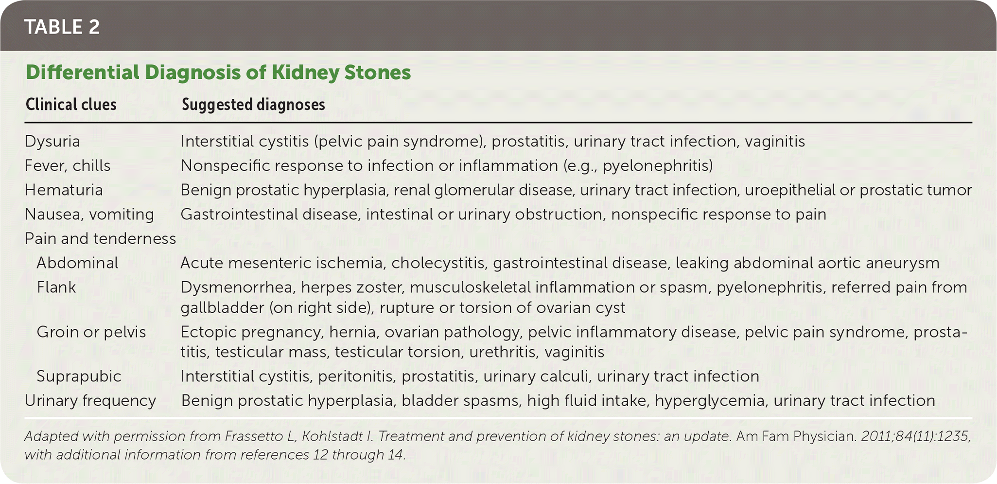
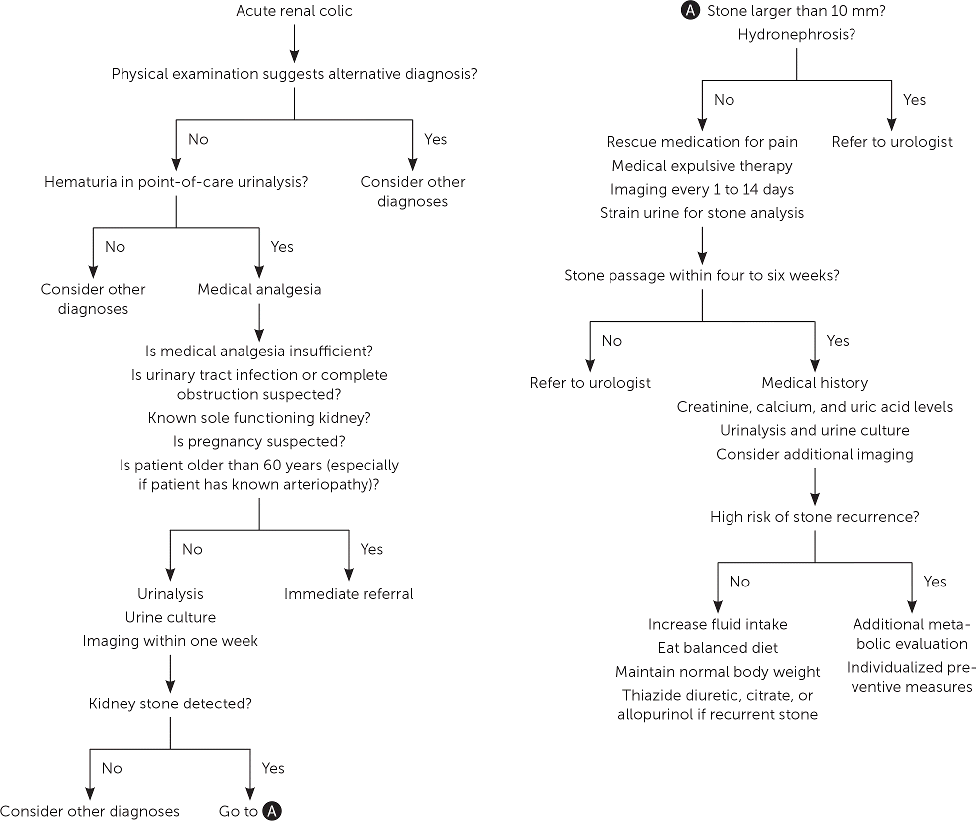
Acute renal colic presents as cramping and intermittent abdominal and flank pain as kidney stones travel down the ureter from the kidney to the bladder.2 Pain is often accompanied by nausea, vomiting, and malaise; fever and chills may also be present.2 Similarity with a previous episode should increase confidence in the diagnosis, although the value of personal or family history during an episode of renal colic is not known. The physical examination should be directed toward excluding differential diagnoses (e.g., urinary tract infection, musculoskeletal inflammation or spasm, ectopic pregnancy, testicular torsion, malignancy; Table 2).2,12–14 The initial workup of a patient with suspected kidney stones in the primary care setting should include point-of-care urinalysis to detect blood, because hematuria helps confirm the diagnosis2,5,13,15 (Figure 1).

| Clinical clues | Suggested diagnoses | |
|---|---|---|
| Dysuria | Interstitial cystitis (pelvic pain syndrome), prostatitis, urinary tract infection, vaginitis | |
| Fever, chills | Nonspecific response to infection or inflammation (e.g., pyelonephritis) | |
| Hematuria | Benign prostatic hyperplasia, renal glomerular disease, urinary tract infection, uroepithelial or prostatic tumor | |
| Nausea, vomiting | Gastrointestinal disease, intestinal or urinary obstruction, nonspecific response to pain | |
| Pain and tenderness | ||
| Acute mesenteric ischemia, cholecystitis, gastrointestinal disease, leaking abdominal aortic aneurysm | |
| Dysmenorrhea, herpes zoster, musculoskeletal inflammation or spasm, pyelonephritis, referred pain from gallbladder (on right side), rupture or torsion of ovarian cyst | |
| Ectopic pregnancy, hernia, ovarian pathology, pelvic inflammatory disease, pelvic pain syndrome, prostatitis, testicular mass, testicular torsion, urethritis, vaginitis | |
| Interstitial cystitis, peritonitis, prostatitis, urinary calculi, urinary tract infection | |
| Urinary frequency | Benign prostatic hyperplasia, bladder spasms, high fluid intake, hyperglycemia, urinary tract infection | |

ACUTE MANAGEMENT
Pain relief is the priority in the acute management of renal colic.5,13 Nonsteroidal anti-inflammatory drugs (e.g., ketorolac, 30 to 60 mg intramuscularly) are more effective and have fewer adverse effects than opioids.5,13,16,17 If an opioid is used, meperidine (Demerol) should be avoided because of the significant risk of nausea and vomiting.17,18 Neither scopolamine nor increased fluid intake alleviates renal colic.16,19
Immediate referral to a urologist or emergency department is warranted when medical analgesia is insufficient; when sepsis is suspected; when anuria, bilateral obstruction, urinary tract infection with renal obstruction, or obstruction of the sole functioning kidney are present; in women who are pregnant or have delayed menstruation (because of the risk of ectopic pregnancy); and in patients who have potential comorbidities or are older than 60 years, especially those with arteriopathy (because of the risk of leaking abdominal aortic aneurysm).5,13,14
DIAGNOSTIC WORKUP
When immediate referral is not indicated, urine culture and urinalysis (if not already done) should be ordered to rule out infection, as well as imaging to confirm the diagnosis of kidney stones and assess for hydronephrosis and stone size and position.2,5,13,15 Although non–contrast-enhanced computed tomography (CT) of the abdomen and pelvis has superior sensitivity and specificity and is commonly performed in the emergency department,5,20–22 first-line ultrasonography has acceptable performance and is more cost-effective.5,13,20 Intravenous urography with plain radiography has limited accuracy and is no longer the preferred diagnostic imaging modality for kidney stones.5 There is no direct evidence for the optimal timing of diagnostic workup for acute renal colic in the primary care setting. However, a 2002 evidence-based consensus review from the United Kingdom recommended that ultrasonography be performed within one week of symptom onset.13 Referral to a urologist for active stone removal is warranted when the stone is larger than 10 mm or if significant hydronephrosis is present.5,13
FOLLOW-UP
Conservative management is indicated if referral is not necessary. Patients should receive pain medication as needed, and follow-up imaging (ultrasonography and possibly plain radiography) should be obtained once within 14 days to monitor evolving stone position and assess for hydronephrosis.5,23 Complete urinary obstruction causes irreversible loss of kidney function, but patients with well-controlled pain and no significant degree of hydronephrosis have only partial obstruction and can be followed for about four to six weeks.5,13,23–26 If the stone does not pass spontaneously, the patient should be referred to a urologist for active stone removal.
Approximately 86% of kidney stones pass spontaneously; this proportion is lower for stones larger than 6 mm (59% vs. 90% for smaller stones).24 Although stones larger than 6 mm in diameter are often removed by urologists,5 these are the stones that have greatest benefit from medical expulsive therapy.27 Medical expulsive therapy with alpha blockers (e.g., tamsulosin [Flomax], 0.4 mg per day; doxazosin [Cardura], 4 mg per day) hastens and increases the likelihood of stone passage, reduces pain, and prevents surgical interventions and hospital admissions.5,27 These medications should be offered to patients with distal ureteral stones 5 to 10 mm in diameter.27 Tamsulosin is the most studied medication, but other alpha blockers seem equally effective.27 Calcium channel blockers (e.g., nifedipine) are less effective and may be no more effective than placebo.28–30 Coadministration of oral corticosteroids or increasing fluid intake does not hasten stone passage or alleviate renal colic.5,19
Further Evaluation in the Subacute Setting
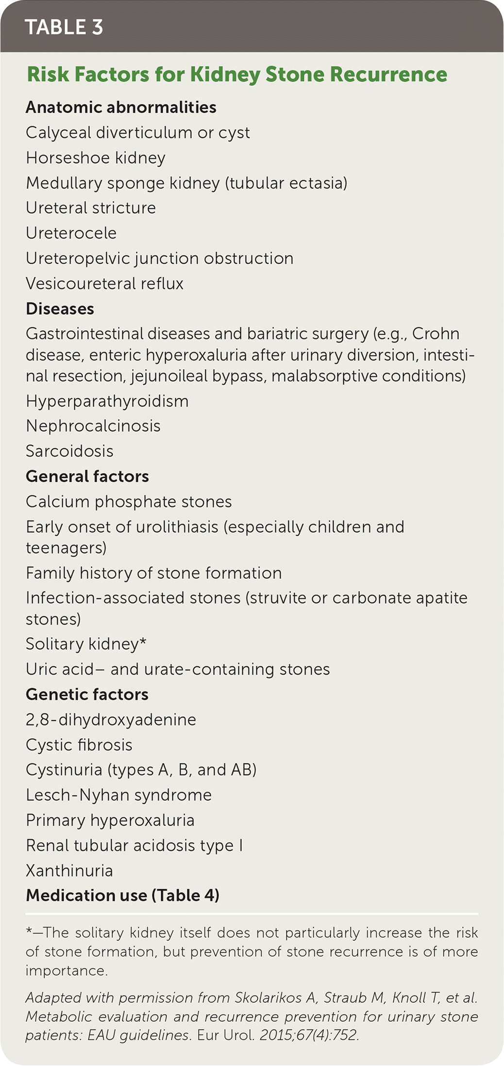
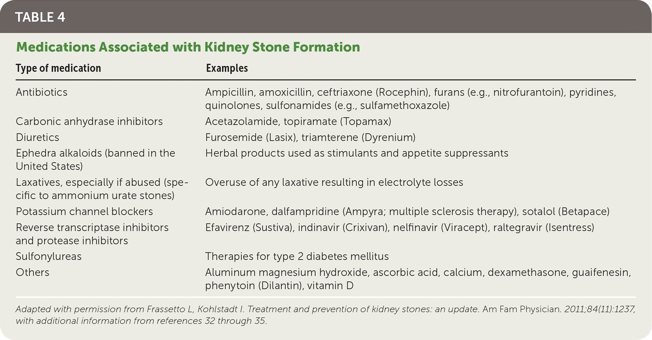
Patients with newly diagnosed kidney stones should receive a basic evaluation consisting of a detailed medical history, serum chemistry, and urinalysis/urine culture. Patients at risk of stone recurrence (Table 331 and Table 42,32–35) should be referred for additional metabolic testing (e.g., 24-hour urine collection for total volume, pH, and calcium oxalate, uric acid, citrate, sodium, potassium, and creatinine levels) and individualized preventive measures.15,31 The medical history should review the stone history (including family history of kidney stones), diet, current medications, and conditions associated with an increased risk of kidney stones.2,15,31–34

|

| Type of medication | Examples |
|---|---|
| Antibiotics | Ampicillin, amoxicillin, ceftriaxone (Rocephin), furans (e.g., nitrofurantoin), pyridines, quinolones, sulfonamides (e.g., sulfamethoxazole) |
| Carbonic anhydrase inhibitors | Acetazolamide, topiramate (Topamax) |
| Diuretics | Furosemide (Lasix), triamterene (Dyrenium) |
| Ephedra alkaloids (banned in the United States) | Herbal products used as stimulants and appetite suppressants |
| Laxatives, especially if abused (specific to ammonium urate stones) | Overuse of any laxative resulting in electrolyte losses |
| Potassium channel blockers | Amiodarone, dalfampridine (Ampyra; multiple sclerosis therapy), sotalol (Betapace) |
| Reverse transcriptase inhibitors and protease inhibitors | Efavirenz (Sustiva), indinavir (Crixivan), nelfinavir (Viracept), raltegravir (Isentress) |
| Sulfonylureas | Therapies for type 2 diabetes mellitus |
| Others | Aluminum magnesium hydroxide, ascorbic acid, calcium, dexamethasone, guaifenesin, phenytoin (Dilantin), vitamin D |
The patient should be instructed to strain his or her urine to catch the stone, then send the stone in a urine specimen cup or a clean, dry container for analysis; non–calcium oxalate stones require additional metabolic testing.15,31 Recurrent stones should also be considered for analysis because their composition may differ from the initial stone.15,31 When stone analysis is not available, ultrasonography should be ordered to look for renal abnormalities if it was not performed before the stone was passed. Non–contrast-enhanced CT should be considered if residual stone is suspected; this modality may help identify stone composition.31
Basic laboratory evaluations include creatinine (for renal function), ionized calcium (for hyperparathyroidism), and uric acid (for hyperuricemia); parathyroid hormone should be measured only if the serum calcium level is high.15,31 If a stone was not retrieved for analysis, additional tests should be considered: urine pH (for nephrocalcinosis and other metabolic abnormalities), microscopy of sediment from morning urine (for urine crystals that may suggest stone composition), and a test for cystinuria (especially in children because it is an inherited metabolic disorder).31
Special Considerations
ASYMPTOMATIC KIDNEY STONES
Many kidney stones are asymptomatic and found on imaging; each year, 10% to 25% become symptomatic or require intervention.5 Conservative management is an option for adults who are healthy, unfit for surgery, or pregnant, and who have access to health care and can adhere to active surveillance (imaging after six months, then annually).5,36 The patient should be referred for stone removal if symptoms, obstruction, or recurrent infection develops, or if the stone grows larger.5,36 Stone removal should be considered if the patient prefers removal to conservative management; plans to conceive in the near future; has calyceal diverticular stones, stones larger than 10 mm (possibly larger than 4 mm), or renal pathology; or is unsuited for conservative management.36
CHILDREN
Kidney stones are becoming more prevalent in children because of increasing rates of diabetes mellitus, obesity, and hypertension in this population.2–4,9 Increasing age is a risk factor for kidney stones; therefore, adolescents are more likely to form stones than younger children.2 Children with kidney stones are more likely to have a metabolic, neurologic, or congenital urinary system structural abnormality; to have concomitant urinary infection; and to have recurrent stones.2,3,9,31
PREGNANT WOMEN
Urinary stasis, increased glomerular filtration rate, and elevated urine pH affect kidney stone formation in pregnant women. Up to 75% of stones in pregnant women are composed of calcium phosphate, in contrast with other adults, in whom calcium oxalate stones are most common.5 Diagnostic and treatment options are limited during pregnancy because of risk to the fetus.5 Kidney stones may increase the risk of preterm labor and other maternal and fetal complications.37
Prevention
Measures to prevent recurrence of kidney stones include lifestyle modifications, citrate supplementation, and medications.2,15,31,38,39 Lifestyle modifications are the cornerstone of prevention after a first kidney stone in patients with low risk of recurrence, whereas citrate supplementation and medications are reserved for patients with recurrent stones.15,31,38,39 Patients at high risk of stone recurrence should receive preventive measures tailored to the results of the metabolic assessment.
LIFESTYLE MODIFICATIONS
The most important lifestyle modification to prevent recurrent kidney stones is to increase fluid intake to 2.5 to 3 L per day to guarantee diuresis of 2 to 2.5 L per day and a urine specific gravity lower than 1.010.15,31,38–40 Fluids should be consumed throughout the day and should consist of beverages with a neutral pH.31 Collection of urine over 24 hours may be necessary to ensure that the diuresis target is met. Decreasing intake of carbonated drinks, especially those acidified with phosphoric acid (e.g., colas), further reduces risk of stone recurrence.38,39
Overall, a balanced diet is ideal for preventing stone recurrence.15,31 The diet should be high in fiber and vegetables, with normal calcium content (1.0 to 1.2 g per day) and limited sodium (4 to 5 g per day) and animal protein (0.8 to 1.0 g per kg per day).15,31 Patients who are obese or over-weight should pursue a normal body weight through dietary modification and increased physical activity.2,15,31 Although there is limited evidence to support lifestyle modifications for the prevention of kidney stone recurrence, these changes are important for preventing comorbidities.
CITRATE SUPPLEMENTATION AND MEDICATIONS
Thiazide diuretics, allopurinol, and citrate supplementation are effective in preventing calcium stones that recur despite lifestyle modification, even in the absence of hyperuricemia, urinary acidosis, hypocitraturia, or hyperuricosuria.15,31,38,39,41 The effectiveness of thiazide diuretics has been documented only with high dosages (e.g., hydrochlorothiazide, 50 mg per day; chlorthalidone, 25 to 50 mg per day; indapamide, 2.5 mg per day); lower dosages have fewer adverse effects, but their effectiveness is unknown.38,39
Allopurinol should be started at 100 mg once per day and increased gradually to 100 mg three times per day.31 There is no evidence that combination therapy with thiazide diuretics or alkaline citrates is more effective than monotherapy.15,31,38,39 Allopurinol is one of the mainstays of therapy for patients with calcium stones, but most patients with uric acid stones have acidic urine that requires treatment with alkaline citrates.15,31
Citrate supplementation is used not only for calcium stones, but also for uric acid (urine pH target 6.0 to 7.5 or greater) and cystine stones (urine pH target of 7.0 to 7.5 or greater).15,31 The preferred salt for supplementation is potassium citrate at a target dosage of 5 to 12 g per day.15,31,38,41 The initial dosage should be 9 g per day, divided into three doses and taken within 30 minutes of meals or a bedtime snack. The most common adverse effects are gastrointestinal symptoms. Potassium citrate supplementation may correct low serum potassium levels caused by thiazide diuretics, but there is no evidence that combination therapy is more effective than monotherapy with either agent.15,31,38,39 Sodium citrate is an alternative for citrate supplementation, but the resulting excretion of sodium and calcium may partially counteract the intended effect.15,31,38 Unsweetened lemonade is a more palatable and less expensive alternative for citrate supplementation. Although there is no direct evidence of its effectiveness in preventing stone recurrence, the dilution of lemon juice in water should help patients meet the recommended fluid intake.42
If medication or citrate supplementation is prescribed, serum potassium levels (for patients taking thiazide diuretics or potassium citrate) and liver enzymes (allopurinol) should be monitored to detect potentially serious adverse effects.15 Potassium levels should be monitored before prescription, within two weeks of prescription, and then every 12 months (earlier if illness occurs or another medication is added).43 There are no recommendations on the frequency of monitoring for hepatotoxicity.
This article updates previous articles on this topic by Frassetto and Kohlstadt2 ; Pietrow and Karellas12 ; Goldfarb and Coe44 ; and Portis and Sundaram.45
Data Sources: We searched PubMed (using PubMed Clinical Queries, ACCESSSS, and Essential Evidence Plus), LILACS (using Virtual Health Library), Essential Evidence, and the Cochrane Database of Systematic Reviews (through PubMed, LILACS, Essential Evidence Plus, and the Cochrane Library) using the key terms kidney calculi, ureterolithiasis, urinary calculi, urolithiasis, or nephrolithiasis. Search dates: November 2017 to December 2018.
