Am Fam Physician. 2021;104(4):online
Author disclosure: No relevant financial affiliations.

Details for This Review
Study Population: 1,860 patients from 10 studies
Efficacy End Points: Successful cannulation on the first attempt (primary); number of attempts, patient satisfaction, and procedure length (secondary)
Harm End Points: Data on harms not reported
Narrative: Peripheral intravenous cannulation is commonly performed in emergency departments and inpatient settings.1 Peripheral intravenous cannulation access can be challenging. One study reported that one out of nine patients in the emergency department met the criteria for difficult access.2 Traditionally, failure to achieve peripheral intravenous cannulation would lead to the placement of a central venous catheter.1 However, the increased availability and use of point-of-care ultrasonography may improve success rates for peripheral intravenous cannulation, with one study reporting that point-of-care ultrasound guidance reduced the need for a central venous catheter by 85% among patients with difficult access.3

| Benefits | Harms |
|---|---|
| 1 in 7 were helped (higher first-attempt cannulation rate) | Not reported |
| 14% increase in successful first-attempt cannulation | |
| 0.3 fewer attempts (mean difference) | |
| 1.5 higher patient satisfaction (mean difference) | |
| No significant difference in procedure length |
This systematic review and meta-analysis included nine randomized controlled trials and one cohort study comprising 1,860 patients and compared ultrasound-guided peripheral intravenous cannulation with the palpation-only technique.4 The ultrasound operators were physicians in three studies, medical students in one study, nurses in four studies, and emergency department technicians in two studies. Most studies used a didactic training session for the operators that ranged from 0.5 to 2 hours. Two studies required that the operators have 10 successful supervised ultrasound-guided peripheral intravenous cannulations. Two other studies required that they have a history of five previously successful ultrasound-guided peripheral intravenous cannulations.
The primary outcome was the rate of successful first attempts as defined by the individual study authors. Secondary outcomes included the number of attempts, patient satisfaction, and procedure length.
Ultrasound-guided peripheral intravenous cannulation was associated with a greater rate of first-attempt success compared with the palpation-only technique (77% vs. 63%; odds ratio = 2.1; 95% CI, 1.65 to 2.7; absolute risk difference = 14%; number needed to treat = 7). Ultrasound-guided peripheral intravenous cannulation was also associated with fewer attempts (standardized mean difference [SMD] = −0.272; 95% CI, −0.539 to −0.004) and higher patient satisfaction (SMD = 1.467; 95% CI, 0.920 to 2.012). There was no difference in the length of the procedure.
Most of the trials did not report on complications or adverse events associated with the procedure; therefore, this outcome was not included in the meta-analysis.
Caveats: There are several limitations to this review. There was significant variation in ultrasound operators (e.g., physician, medical student, nurse, technician) and their experience level in obtaining vascular access. There were also variations in the definitions for procedure length and confirmation of successful first-attempt cannulation. There was limited reporting on complication rates, and most of the data were derived from one large study,5 although this study was deemed at low risk of bias. Many of the other studies were deemed moderate to high risk of bias, and the inclusion of a cohort study may have introduced bias because of the lack of randomization.
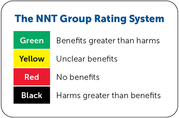
Conclusion: Based on existing evidence, we have assigned a color recommendation of green (benefits greater than harms) for the use of ultrasound guidance for peripheral intravenous cannulation. Further data are needed to better identify which factors are most predictive of success and to determine the impact on procedure length and patient satisfaction.
