
Am Fam Physician. 2022;105(6):671-672
Author disclosure: No relevant financial relationships.
An 11-year-old girl presented for evaluation of multiple pruritic lesions. The lesions began when she was two or three years of age. Over time, they evolved to become fixed and increased in number. The patient had severe pruritus and flushing of her face. Her medical history was significant for frequent headaches, a one-year history of diarrhea, and one episode of difficulty breathing that was associated with exertion and required evaluation by emergency medical services.

Question
A. Atopic dermatitis.
B. Cutaneous mastocytosis.
C. Idiopathic anaphylaxis.
D. Neurofibromatosis 1.
E. Xanthoma.
Discussion
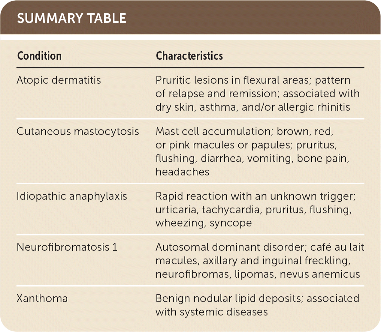
The answer is B: cutaneous mastocytosis. Mastocytosis refers to a heterogeneous group of conditions characterized by excess production and accumulation of mast cells within the skin (cutaneous mastocytosis) or other organs such as the bone marrow, gastrointestinal tract, or skeletal system (systemic mastocytosis).1 Cutaneous mastocytosis accounts for about 90% of cases in children.1 Onset typically occurs before two years of age, but lesions may be present at birth.2 Cutaneous mastocytosis can be categorized into maculopapular cutaneous mastocytosis (formerly known as urticaria pigmentosa; 75% of cases), solitary mastocytoma (20%), and diffuse cutaneous (5%).2,3
In children, manifestations are usually limited to the skin. Systemic symptoms caused by mast cell mediator release can be present even without the infiltration of mast cells into other tissues. The most commonly reported systemic symptom is pruritus, followed by flushing, diarrhea, vomiting, bone pain, and headaches.1–3 Childhood onset of maculopapular cutaneous mastocytosis often presents with brown, red, or pink macules or papules, with asymmetrical and widespread distribution.3,4
Cutaneous mastocytosis is diagnosed clinically and can be confirmed with a skin biopsy.5 The Darier sign (wheal and flare reaction occurring after rubbing a lesion about five times with moderate pressure using a tongue depressor) is a highly specific diagnostic feature.3 Serum tryptase levels should be measured at initial presentation of mastocytosis to rule out systemic involvement and to identify patients at risk of severe mast cell mediator release symptoms.
Atopic dermatitis is the most common inflammatory skin condition in children and manifests as pruritic lesions in flexural areas (e.g., elbows, behind knees, ankles, neck).6 Atopic dermatitis occurs in a pattern of relapse and remission and usually involves a history of dry skin, asthma, and/or allergic rhinitis.
Idiopathic anaphylaxis is a rare but potentially life-threatening rapid reaction that may occur with mastocytosis. Other symptoms, which may involve the skin or respiratory, cardiovascular, or gastrointestinal systems, include urticaria, tachycardia, pruritus, flushing, wheezing, and syncope.7
Neurofibromatosis 1 is a rare autosomal dominant disorder that presents with various cutaneous manifestations in adolescence. The most common cutaneous findings include café au lait macules, axillary and inguinal freckling, neurofibromas, lipomas, and nevus anemicus.8
| Condition | Characteristics |
|---|---|
| Atopic dermatitis | Pruritic lesions in flexural areas; pattern of relapse and remission; associated with dry skin, asthma, and/or allergic rhinitis |
| Cutaneous mastocytosis | Mast cell accumulation; brown, red, or pink macules or papules; pruritus, flushing, diarrhea, vomiting, bone pain, headaches |
| Idiopathic anaphylaxis | Rapid reaction with an unknown trigger; urticaria, tachycardia, pruritus, flushing, wheezing, syncope |
| Neurofibromatosis 1 | Autosomal dominant disorder; café au lait macules, axillary and inguinal freckling, neurofibromas, lipomas, nevus anemicus |
| Xanthoma | Benign nodular lipid deposits; associated with systemic diseases |
