Am Fam Physician. 2023;107(3):305-306
Author disclosure: No relevant financial relationships.
A 52-year-old woman presented with black discoloration of the fingernails (Figure 1). She had no trauma to the area and no associated pain, rash, swelling, or drainage. She had no other symptoms, including shortness of breath, chest pain, or visual disturbances. Her medical history was significant for type 2 diabetes mellitus, hypertension, thrombocytosis, and coronary artery bypass. The patient was taking metformin, glipizide, lisinopril, hydroxyurea, and topiramate.

Her vital signs were normal on physical examination. All fingernails showed black discoloration but no pitting, spooning, or ulceration. The fingertips were warm to the touch and nontender to palpation.
Question
Based on the patient’s history and physical examination findings, which one of the following is the most likely diagnosis?
A. Mees lines.
B. Melanonychia.
C. Onychomycosis.
D. Subungual hematoma.
E. Subungual melanoma.
Discussion
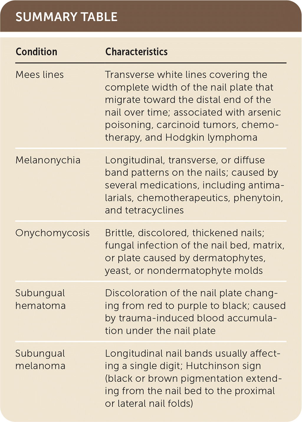
The answer is B: melanonychia. Several medications are known to induce black discoloration of fingernails, including antimalarials, chemotherapeutics (hydroxyurea, cyclophosphamide, fluorouracil), phenytoin, and tetracyclines.1 The discoloration may present as a longitudinal, transverse, or diffuse band pattern. Patients may have coexisting diffuse pigmentation of the skin (melanoderma). Melanonychia can also occur with nutritional deficiencies (folate, vitamin B12), connective tissue diseases (systemic lupus erythematosus, scleroderma), endocrinopathies (Addison disease, Cushing syndrome, hyperthyroidism), or physiologic processes (pregnancy).1
This patient had been treated with hydroxyurea for thrombocytosis. Nail changes caused by antineoplastic agents are not associated with other local or systemic symptoms and are reversible within a few months of discontinuing the causative agent.2 A thorough history and physical examination are essential to exclude other causes of fingernail discoloration.
Mees lines are transverse white lines covering the complete width of the nail plate. They can affect a single nail or multiple nails. A key distinguishing feature is migration toward the distal end of the nail over time because the abnormality is predominantly in the nail plate.3 Mees lines are associated with arsenic poisoning, carcinoid tumors, chemotherapy, and Hodgkin lymphoma.3
Onychomycosis is a fungal infection of the nail bed, matrix, or plate caused by dermatophytes, yeast, or nondermatophyte molds.4 It leads to brittle, discolored, thickened nails. Pain, discomfort, and physical impairment can occur without treatment. Accurate diagnosis using potassium hydroxide preparation with direct microscopy is recommended before starting treatment.4 A combination of nail trimming, debridement, and pharmacotherapy is the most effective treatment strategy.4
Subungual hematomas are caused by trauma to the nail bed. The nail discoloration changes from red to purple to black as the blood clots. Treatment is predominantly supportive, but clot evacuation and nail removal are options if the hematoma causes severe pain.3
Subungual melanoma presents as longitudinal nail bands and is unlikely to affect multiple digits. Hutchinson sign, an indicator of subungual melanoma, is black or brown pigmentation extending from the nail bed to the proximal or lateral nail folds.3

| Condition | Characteristics |
|---|---|
| Mees lines | Transverse white lines covering the complete width of the nail plate that migrate toward the distal end of the nail over time; associated with arsenic poisoning, carcinoid tumors, chemotherapy, and Hodgkin lymphoma |
| Melanonychia | Longitudinal, transverse, or diffuse band patterns on the nails; caused by several medications, including antimalarials, chemotherapeutics, phenytoin, and tetracyclines |
| Onychomycosis | Brittle, discolored, thickened nails; fungal infection of the nail bed, matrix, or plate caused by dermatophytes, yeast, or nondermatophyte molds |
| Subungual hematoma | Discoloration of the nail plate changing from red to purple to black; caused by trauma-induced blood accumulation under the nail plate |
| Subungual melanoma | Longitudinal nail bands usually affecting a single digit; Hutchinson sign (black or brown pigmentation extending from the nail bed to the proximal or lateral nail folds) |
