Am Fam Physician. 2023;107(4):427-428
Author disclosure: No relevant financial relationships.
A 73-year-old woman presented with a rapidly growing, painful sore on her thigh. The lesion appeared spontaneously two weeks earlier without any identified triggers. It did not drain, bleed, or itch. The patient had no systemic symptoms, including fever, chills, or weight loss. She had a history of recurrent pus-filled boils that required incision and drainage.
Physical examination revealed a sharply demarcated, firm, erythematous lesion on her right, medial thigh that was nonfluctuant and had a punctate central plug (Figure 1). It was 0.7 × 0.6 cm in size and tender to palpation around the edges. Her vital signs were normal. A shave biopsy was performed.

Question
Based on the patient's history and physical examination findings, which one of the following is the most likely diagnosis?
A. Cutaneous squamous cell carcinoma.
B. Folliculitis.
C. Keratoacanthoma.
D. Nodular basal cell carcinoma.
E. Skin abscess.
Discussion
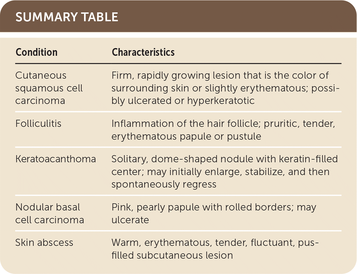
The answer is A: cutaneous squamous cell carcinoma, which was confirmed on biopsy. Cutaneous squamous cell carcinoma is a malignant neoplasm of the skin that typically arises from actinic keratosis. Cutaneous squamous cell carcinoma classically presents as a firm, rapidly growing lesion that is the color of the surrounding skin or slightly erythematous. It can present as central ulceration or hyperkeratosis, as in this patient.1 Smoking and exposure to ultraviolet radiation are predisposing factors. The head and neck are the most common locations for these lesions.2 Evaluation of suspicious lesions should begin with a full-body inspection, followed by dermatoscopy and biopsy. Early detection and removal decrease the likelihood of metastasis and significantly improve prognosis.3
Folliculitis is inflammation of a hair follicle that presents as a pruritic, tender, erythematous papule or pustule.4 The cause can be infectious or noninfectious. Diagnosis is clinical, but lesions can be further evaluated using Gram stain, culture, or biopsy. Treatment includes hot compresses, antibacterial ointment, and antibiotics.5
Keratoacanthomas are keratin-filled epithelial tumors that clinically and histologically resemble cutaneous squamous cell carcinoma.6 They present as a solitary, dome-shaped nodule primarily on sun-exposed areas. They are distinguished from carcinoma by an unpredictable progression. Although some grow steadily, others initially enlarge, stabilize, and then spontaneously regress.7
Nodular basal cell carcinoma, the most common basal cell variant, is a malignant skin lesion that originates from the basal layer of the epidermis.8 Although it can mimic squamous cell carcinoma, its defining characteristics include a pearly texture, pink coloration, and rolled borders. Nodular basal cell carcinomas occasionally ulcerate, mimicking the crusted, punctate opening seen in this patient.9 Skin abscesses are collections of pus in the dermis or deeper subcutaneous tissue, secondary to infection. They typically present as warm, erythematous, tender, fluctuant lesions, occasionally causing systemic symptoms, including fever, chills, and lymphadenopathy. Abscesses may spontaneously drain or may require incision and drainage for full resolution.10

| Condition | Characteristics |
|---|---|
| Cutaneous squamous cell carcinoma | Firm, rapidly growing lesion that is the color of surrounding skin or slightly erythematous; possibly ulcerated or hyperkeratotic |
| Folliculitis | Inflammation of the hair follicle; pruritic, tender, erythematous papule or pustule |
| Keratoacanthoma | Solitary, dome-shaped nodule with keratin-filled center; may initially enlarge, stabilize, and then spontaneously regress |
| Nodular basal cell carcinoma | Pink, pearly papule with rolled borders; may ulcerate |
| Skin abscess | Warm, erythematous, tender, fluctuant, pus-filled subcutaneous lesion |
