Am Fam Physician. 2024;110(5):531-532
Author disclosure: No relevant financial relationships.
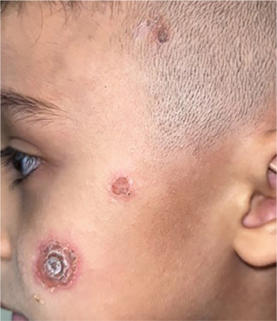
A 5-year-old boy presented with lesions on his left cheek and temporal area that had appeared a few days earlier. Each lesion started as an erythematous papule and evolved into a coin-sized ulcerated plaque. The plaques featured brown, central adherent crusts and yellow-brown, dried marginal exudates with surrounding erythema (Figure 1).

The patient did not have a history of insect or arthropod bites, and he did not report pain, pruritus, or associated fever or chills. Fusidic acid (a topical antibacterial) was applied to the lesions, but this did not lead to improvement. The patient was then prescribed cephalexin (50 mg/kg per day). One week later, the lesions had improved significantly.
QUESTION
Based on the patient's history and physical examination, which one of the following is the most likely diagnosis?
A. Ecthyma.
B. Ecthyma gangrenosum.
C. Impetigo.
D. Pyoderma gangrenosum.
DISCUSSION
The answer is A: ecthyma. This ulcerative bacterial skin infection is caused by group A streptococci and is often associated with secondary staphylococcal infection. Ecthyma typically begins superficially and extends into the dermis. It can be referred to as a deeper form of impetigo. However, impetigo affects only the stratum corneum, whereas ecthyma extends into the dermis and results in scarring.1
Subscribe
From $180- Immediate, unlimited access to all AFP content
- More than 125 CME credits/year
- AAFP app access
- Print delivery available
Issue Access
$59.95- Immediate, unlimited access to this issue's content
- CME credits
- AAFP app access
- Print delivery available
