Am Fam Physician. 2024;110(6):635-636
Author disclosure: No relevant financial relationships.
A 68-year-old man presented with black discoloration of his left great toe and toenail that had been present for more than 1 year. The toe was not painful. Treatment with antifungal spray did not lead to improvement. The patient did not have a history of injury or trauma to the toe.
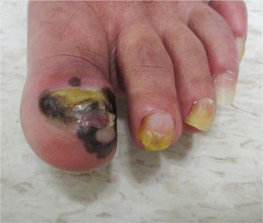
Physical examination revealed a large, irregularly pigmented black and brown plaque of the subungual skin from the left great toe to the proximal and medial nail folds (Figure 1). The toenail was almost completely destroyed, and a large pink nodule with purulent discharge was present in the nail bed. Pinprick sensation was decreased on the plantar surface of the first and second toes.

QUESTION
Based on the patient's history and physical examination, which one of the following is the most likely diagnosis?
A. Diabetic foot ulcer.
B. Longitudinal melanonychia striata.
C. Onychomycosis.
D. Pyogenic granuloma.
E. Subungual melanoma.
DISCUSSION
The answer is E: subungual melanoma. Sixty-five percent of cases present as a longitudinal band of nail discoloration with proximal widening and irregular side borders. The nail plate may also thicken or split.1 Patients may present with diffuse melanonychia, nail dystrophy, nail bed lesions or ulceration, subungual masses, osseous involvement, and lack of gross pigmentation. Hutchinson sign (brown-black pigmentation of subungual skin extending to proximal and lateral nail folds) is associated with subungual melanoma, with sensitivity of 42% and specificity of 96%.2,3 However, it may also occur with subungual hematoma, fungal infection, and drug-induced dyschromia.4,5
Subscribe
From $180- Immediate, unlimited access to all AFP content
- More than 125 CME credits/year
- AAFP app access
- Print delivery available
Issue Access
$59.95- Immediate, unlimited access to this issue's content
- CME credits
- AAFP app access
- Print delivery available
