
Am Fam Physician. 2025;111(3):277-278
Author disclosure: No relevant financial relationships.
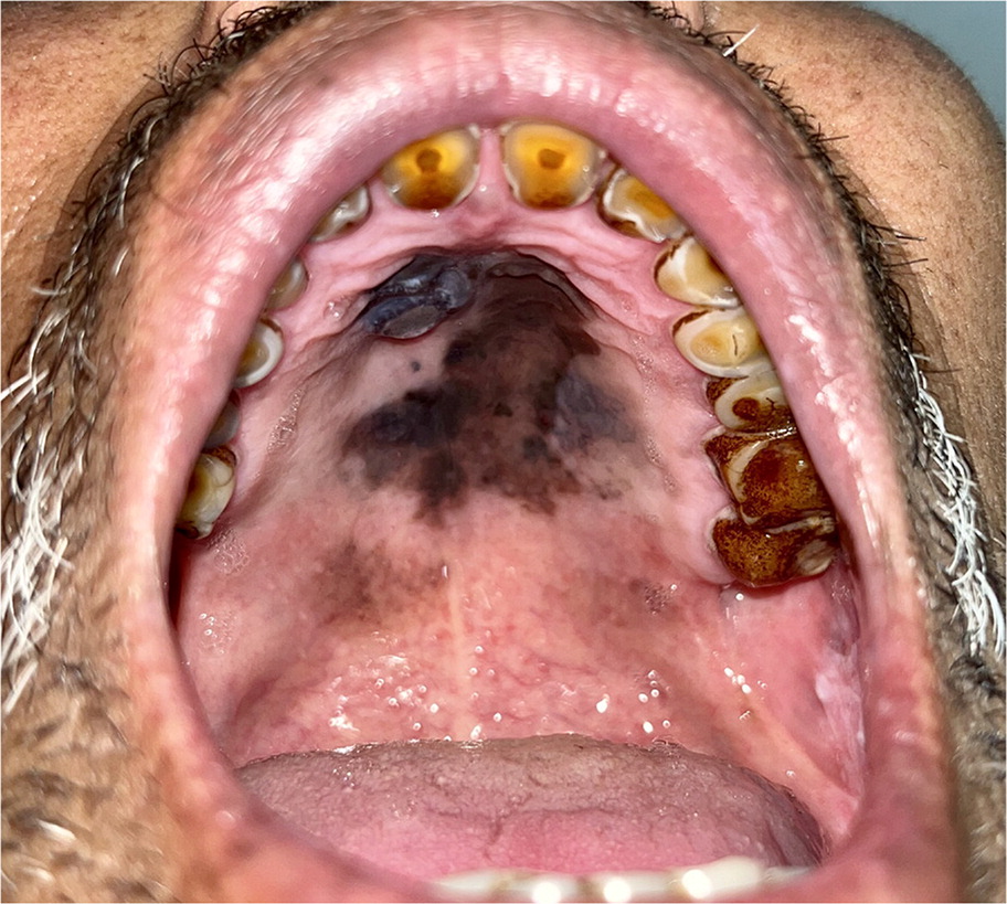
A 59-year-old man presented with a brown-black lesion on the hard palate. The lesion was associated with pain and caused difficulty eating. The lesion started as a small, black-pigmented area 2 years ago and grew gradually. The patient was a 10-pack-year smoker.
Examination of the oral cavity revealed poor oral hygiene and nicotine staining of the teeth. A sharply demarcated, variably pigmented lesion with superimposed nodularity and irregular borders covered a large area of the hard palate (Figure 1). The patient’s upper cervical lymph nodes were bilaterally enlarged (2 cm in diameter) and were mobile, firm, and nontender.
QUESTION
Based on the patient’s history and physical examination, which one of the following is the most likely diagnosis?
A. Drug-induced melanosis.
B. Melanocytic nevus.
C. Melanotic macule.
D. Palatal melanoma.
E. Smoker’s melanosis.
DISCUSSION
The answer is D: palatal melanoma. Oral melanoma usually presents with three key features: a brown-black pigmented plaque, light-brown macular area, and central area with nodularity. Smoking has an unclear relationship with the development of head and neck mucosal melanoma, but it is correlated with increased melanocyte proliferation in the oral mucosa, increasing the risk of pigmented oral lesions.1
Subscribe
From $180- Immediate, unlimited access to all AFP content
- More than 125 CME credits/year
- AAFP app access
- Print delivery available
Issue Access
$59.95- Immediate, unlimited access to this issue's content
- CME credits
- AAFP app access
- Print delivery available
