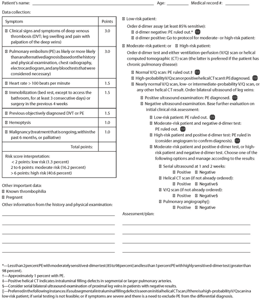
This evidence-based encounter form will help you rule in or rule out PE.
Fam Pract Manag. 2004;11(2):61-63
Mr. Smith is a 62-year-old patient who complains of increasing shortness of breath over the past 24 hours. He has no swelling of the legs, no pain in the calves on palpation and no cough, fever or other symptoms consistent with an alternate diagnosis such as pneumonia. He denies hemoptysis and has no history of malignancy or previous venous thromboembolism. However, he did recently drive eight hours from Orlando with his grandchildren. His heart rate is 104 beats per minute. What is the probability that he has pulmonary embolism (PE) and, based on his risk level, what tests should you run in order to rule in or rule out PE?
What is the patient’s risk?
One of the first steps in diagnosing PE, or any other condition, is to assess the patient’s risk level for the condition based on his or her signs and symptoms. Because individual signs and symptoms are often not accurate enough to estimate a patient’s risk level, combining them into a “clinical decision rule” can be very helpful.
A number of authors have developed and validated clinical decision rules to determine the likelihood of PE. One of the most carefully tested rules, developed by Wells and colleagues,1 requires only a careful history and physical examination. (For more information on the Wells rule, seeAmerican Family Physician, Jan. 15, 2004, page 367.) The PE encounter form uses the seven elements of the Wells rule in the “symptom” section to help you estimate a patient’s risk of PE and then guides you through a diagnostic protocol. An experienced clinician’s estimate of the likelihood of PE without using any clinical decision rule is also reasonably accurate.2,3
If the risk assessment based on a clinical decision rule differs from your “gut” clinical assessment, it seems prudent to rely on the assessment that places the patient in the highest risk group. For example, if the Wells rule places a patient in the low-risk group, but you have a higher index of suspicion based on your global assessment of the patient, consider the risk moderate.
Pulmonary Embolism Encounter Form

Developed by Mark H. Ebell, MD, MS. Copyright (c) 2004 American Academy of Family Physicians. Physicians may photocopy or adapt for use in their own practices; all other rights reserved. "Diagnosing Pulmonary Embolism." Ebell MH. Family Practice Management. February 2004:61-63; https://www.aafp.org/fpm/20040200/61diag.html.
What tests should I order?
Once you have determined a patient’s risk for PE, the next questions are “What tests should I order to rule in or rule out the condition?” and “How should I interpret them?”
One of the strengths of an evidence-based approach to diagnosis is that it allows you to tailor the diagnostic strategy to the patient. Rather than taking a “one size fits all” approach that over-investigates patients who are at low risk and may miss disease in patients who are at high risk, you can use the information from your clinical evaluation to guide the selection of tests and their interpretation. Several groups have developed and validated protocols for the diagnosis of PE that rely on the clinical assessment, D-dimer test, ventilation-perfusion (V/Q) scan, ultrasound of the proximal leg veins and helical computed tomography (CT) scanning.1,3,4,5
The recommended protocol shown in the encounter form is based on a validated protocol developed by Wells and modified by Kearon to add the option of helical CT instead of V/Q scan. Unlike protocols proposed by Musset and Perrier, this one does not require angiography except as an option in a small percentage of patients with indeterminate findings. The protocol is based on the finding that patients with a low clinical probability and a negative noninvasive test almost certainly do not have PE, while patients with a high clinical probability and a positive noninvasive test almost certainly do. Those with an intermediate clinical probability or an indeterminate noninvasive test require further rounds of testing and closer follow-up. It is also important for clinicians to understand that many of the tests in question are “asymmetrical.” That is, they are helpful at ruling in disease when positive or ruling out disease when negative, but not both. For example, the D-dimer test is quite sensitive; when negative in a patient with low risk, it is very good at ruling out PE. However, a positive D-dimer does not rule in the diagnosis; it indicates further confirmatory testing. Conversely, helical CT is very good at ruling in disease when intraluminal filling defects in segmental or larger pulmonary arteries are seen, but it is unhelpful if results are normal or indeterminate. Patients with non-diagnostic V/Q scans and helical CT scans need further testing with ultrasonography of the leg veins to help diagnose or exclude PE.
This protocol applies to adult patients presenting with new or worsening shortness of breath or chest pain to the emergency department or outpatient setting. Patients whose symptoms last for more than 30 days, those with no symptoms for the three days prior to presentation, those with recent anticoagulation, inpatients, pregnant women, patients with suspected thrombosis of an upper extremity vein, and children were excluded from the validation studies and should not be evaluated using this algorithm. Critically ill patients and those with a limited cardiovascular reserve may require more extensive evaluation, since the consequences of a small missed PE are greater in this group. The D-dimer test is much less specific in hospitalized patients, and the V/Q scan is often associated with nondiagnostic results in patients with chronic pulmonary disease.
POINT-OF-CARE SERIES
This article is part of a series that offers evidence-based tools to assist family physicians in improving their decision making at the point of care. The series is produced in partnership with American Family Physician. A related article, which also includes the pulmonary embolism encounter form, appears in the Feb. 1, 2004, issue of AFP, pages 599-601.
The diagnosis
In the case of Mr. Smith, our fictitious patient, the probability of PE is moderate (he has 4.5 points because of his heart rate and no alternative diagnosis). Following the protocol, you would send him for a stat D-dimer test and a helical CT scan. If the D-dimer turned out to be negative, this would not be enough to exclude PE, given the intermediate clinical risk. If we assume that the helical CT revealed an intraluminal filling defect in the segmental arteries, it would confirm the diagnosis of PE. Had the helical CT shown only a subsegmental intraluminal filling defect, further testing (beginning with an ultrasound of the leg veins) would have been indicated to help establish the diagnosis.