Am Fam Physician. 2022;106(4):453-454
Author disclosure: No relevant financial relationships.
A 52-year-old woman presented with a moderately pruritic rash on her right leg that developed several months earlier. She did not have blisters, cough, sore throat, chills, diarrhea, fevers, or joint aches. She had no recent infections or household contacts with a similar rash. She was not using any new medications or personal care products. No improvement was noted after two weeks of treatment with oral prednisone.
Physical examination revealed a purple papular eruption extending in a streak pattern from the medial aspect of the right thigh to the posteromedial aspect of the right calf (Figure 1). Close examination revealed fine white lines within purple plaques. Vital signs were normal.

Question
Based on the patient's history and physical examination findings, which one of the following is the most likely diagnosis?
A. Follicular keratosis.
B. Inflammatory linear verrucous epidermal nevus.
C. Lichen striatus.
D. Linear lichen planus.
E. Linear psoriasis.
Discussion
The answer is D: linear lichen planus. The prevalence of lichen planus is 0.5% to 1%. Linear lichen planus is a rare form of the condition (less than 1% of cases) that usually presents as unilateral pruritic, violaceous, flat-topped papules in a linear distribution on the limbs.1 Fine white lines on top of the plaques (Wickham striae) may be seen. The underlying cause of lichen planus is unknown, although the linear form may be associated with a dermatomal pattern or the Koebner phenomenon at the site of trauma.2 Classic lesions present with the four P's: purple, pruritus, polygonal, and papules/plaques.
In this patient, punch biopsy findings were consistent with linear lichen planus with no evidence of dysplasia or malignancy. Linear lichen planus is histologically identical to classic lichen planus, including hyperkeratosis, focal hypergranulosis, irregular acanthosis with a sawtooth appearance, vacuolar changes in the basal cell layer, and a dense, bandlike lymphocytic infiltrate at the dermal-epidermal junction.3
Follicular keratosis (also called Darier disease) is characterized by reddish-brown, keratotic papules in seborrheic areas, often with a linear distribution. Histologic evaluation demonstrates acantholysis and dyskeratosis.3
Inflammatory linear verrucous epidermal nevus typically presents at birth or in early childhood as scaly, pruritic plaques or papular psoriasiform lesions along Blaschko lines. Histologic evaluation demonstrates hypergranulosis and parakeratosis alternating with hypogranulosis and orthokeratosis.4
Linear psoriasis is a rare form of psoriasis that presents as erythematous, scaly plaques in a linear distribution. Histologic evaluation demonstrates hyperkeratosis, parakeratosis, and hypogranulosis.7

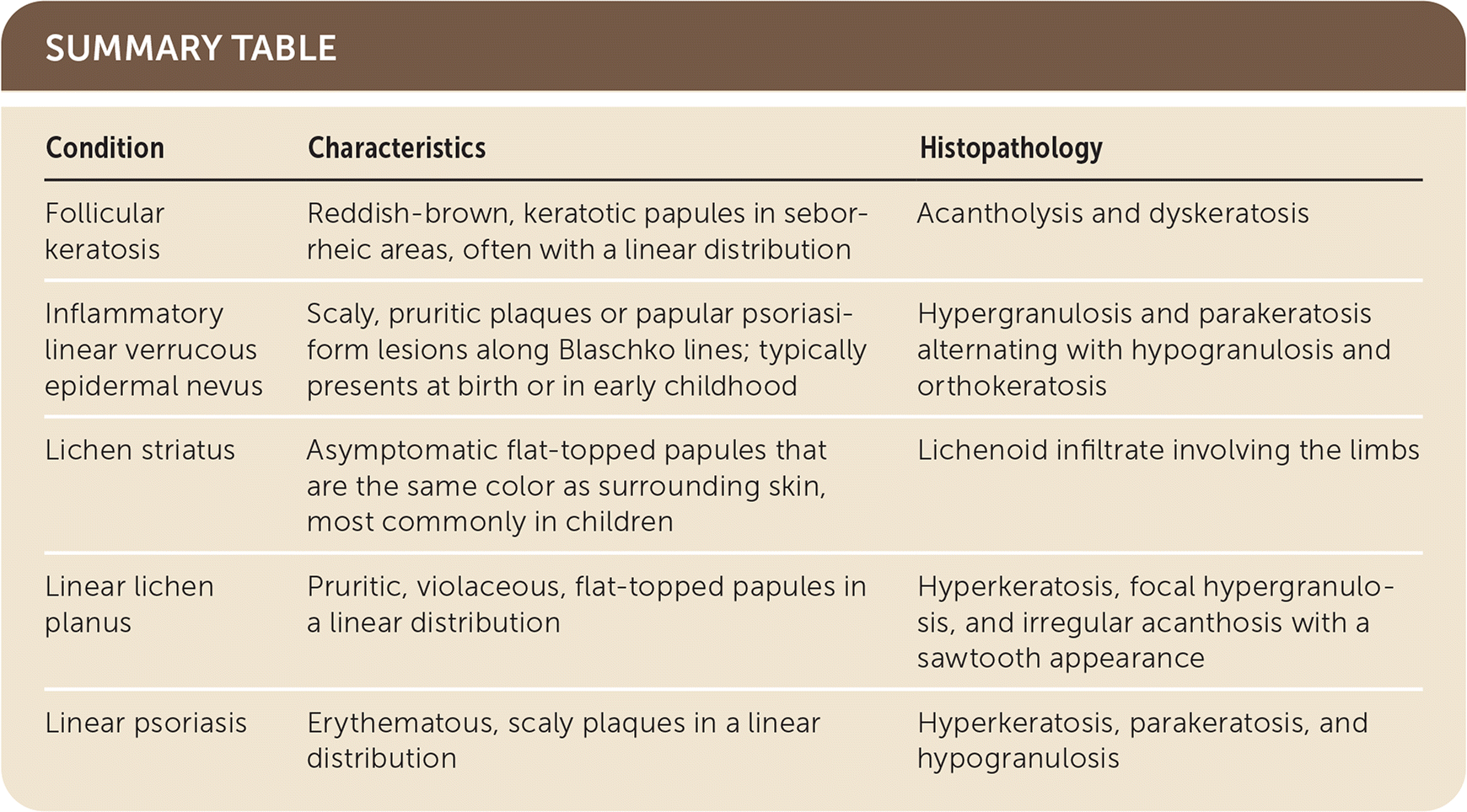
| Condition | Characteristics | Histopathology |
|---|---|---|
| Follicular keratosis | Reddish-brown, keratotic papules in seborrheic areas, often with a linear distribution | Acantholysis and dyskeratosis |
| Inflammatory linear verrucous epidermal nevus | Scaly, pruritic plaques or papular psoriasiform lesions along Blaschko lines; typically presents at birth or in early childhood | Hypergranulosis and parakeratosis alternating with hypogranulosis and orthokeratosis |
| Lichen striatus | Asymptomatic flat-topped papules that are the same color as surrounding skin, most commonly in children | Lichenoid infiltrate involving the limbs |
| Linear lichen planus | Pruritic, violaceous, flat-topped papules in a linear distribution | Hyperkeratosis, focal hypergranulosis, and irregular acanthosis with a sawtooth appearance |
| Linear psoriasis | Erythematous, scaly plaques in a linear distribution | Hyperkeratosis, parakeratosis, and hypogranulosis |
