Am Fam Physician. 2022;106(5):573-574
Author disclosure: No relevant financial relationships.
A 30-year-old woman presented with bilateral wrist pain that began about eight months earlier. The pain progressed in severity and was associated with tingling, numbness, weakness, and aching in both forearms. The patient used a thumb spica splint at night with occasional but inconsistent relief. The pain did not improve after treatment with oral and topical analgesics. After pain onset, areas of skin discoloration developed on both wrists. The patient had no occupational exposures and had not used any new skin products. She had no relevant family history.
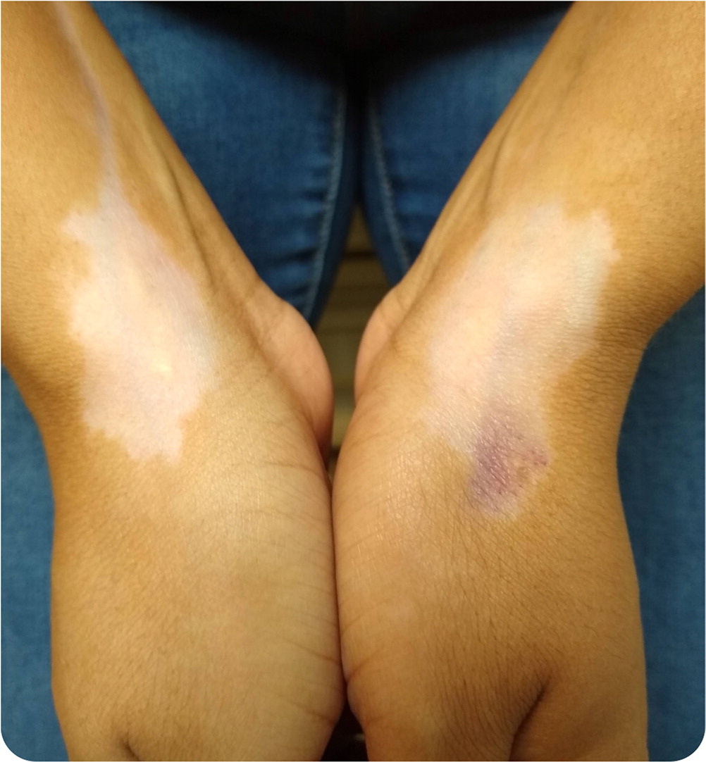
Physical examination revealed bilateral 3- to 4-cm nonraised, nonscaly, hypopigmented patches with irregular borders extending over the first dorsal compartments (Figure 1). There was a 1-cm bruise within the hypopigmented patch on the left side. The lesions were not pruritic or painful. The patient had no other skin rashes or lesions.

Question
Based on the patient’s history and physical examination findings, which one of the following is the most likely diagnosis?
A. Chemical leukoderma.
B. Intra-articular steroid injection hypopigmentation.
C. Pityriasis versicolor.
D. Vitiligo.
Discussion
The answer is B: intra-articular steroid injection hypopigmentation. Hypopigmentation is a common condition associated with decreased melanin production in the affected area compared with surrounding skin.1,2 Areas of hypopigmentation are more prominent in darker skin tones and can be upsetting for patients, especially when located on exposed areas of the body.3
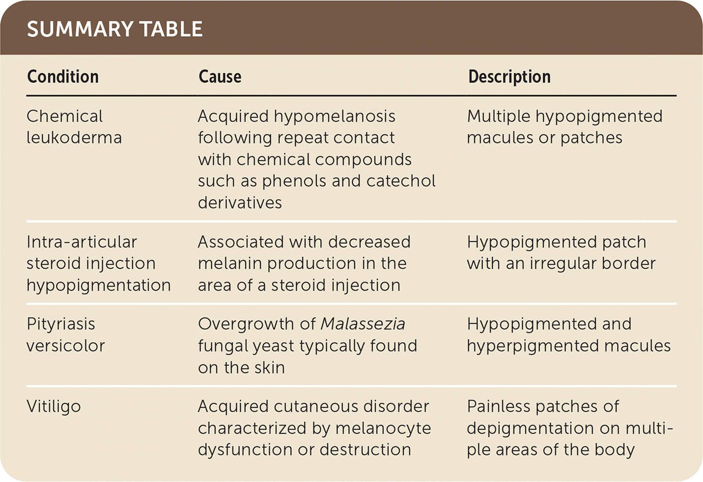
The diagnosis of intra-articular steroid injection hypopigmentation is based on the temporal association between local injection and rash onset. The rash presents as a hypopigmented patch with an irregular border. Triamcinolone injections are routinely used to relieve joint inflammation. These injections are typically well tolerated. However, potential adverse effects of steroid injections include local atrophy, hypopigmentation, hyperpigmentation, and calcification at the injection site.4 Hypopigmentation is rare, occurring in 1% to 4% of patients receiving steroid injections, and is more common following injections of small or superficial joints.5
The patient was treated with intra-articular triamcinolone injection to decrease bilateral wrist inflammation from de Quervain tenosynovitis. This condition is often caused by chronic wrist overuse that leads to pain and inflammation, although its inflammatory etiology has recently been questioned.3 Triamcinolone has high density and low solubility when administered via intrasheath injection, which can lead to drug aggregation and the development of adverse effects. Most patients improve over time, but it may take up to one year.
Chemical leukoderma is an acquired hypomelanosis occurring after repeat contact with chemical compounds such as phenols and catechol derivatives, which exhibit selective melanocytotoxicity that mimics clinically idiopathic vitiligo and other acquired hypopigmentation disorders.6 Lesions appear as multiple hypopigmented macules or patches incited by exposure to agents such as household disinfectants and detergents, hydroquinone, p-phenylenediamine used in hair dyes, cosmetic products, arsenic, mercurials, corticosteroids, chloroquine, and tretinoin. 7 A history of repeated exposures confirms the diagnosis.
Pityriasis versicolor is a benign skin condition characterized by overgrowth of Malassezia fungal yeast typically found on the skin. Lesions comprise both hypopigmented and hyper-pigmented macules, with a varying clinical course of relapsing to chronic infection.8 Hypopigmentation due to pityriasis versicolor is usually seen on the back, chest, and abdomen and is pruritic.
Vitiligo is an acquired cutaneous disorder characterized by melanocyte dysfunction or destruction, which results in painless patches of depigmentation on multiple areas of the body. The etiology of this disorder is unclear, although genetics and autoimmune pathogenesis such as Addison disease, psoriasis, type 1 diabetes mellitus, and pernicious anemia are often associated with vitiligo.9

| Condition | Cause | Description |
|---|---|---|
| Chemical leukoderma | Acquired hypomelanosis following repeat contact with chemical compounds such as phenols and catechol derivatives | Multiple hypopigmented macules or patches |
| Intra-articular steroid injection hypopigmentation | Associated with decreased melanin production in the area of a steroid injection | Hypopigmented patch with an irregular border |
| Pityriasis versicolor | Overgrowth of Malassezia fungal yeast typically found on the skin | Hypopigmented and hyperpigmented macules |
| Vitiligo | Acquired cutaneous disorder characterized by melanocyte dysfunction or destruction | Painless patches of depigmentation on multiple areas of the body |
