Am Fam Physician. 2023;107(1):85-86
Author disclosure: No relevant financial relationships.
A 63-year-old man presented with rapidly spreading pruritic blisters on his upper and lower extremities, chest, and back. The blisters initially appeared three to four weeks earlier on the dorsal aspect of both feet and hands. The large blisters waxed and waned and were filled with clear fluid. A dermatologist had recently biopsied the lesions and prescribed clobetasol cream and oral doxycycline. Three days after starting the medications, the lesions rapidly progressed to include his back and chest. The patient did not have pain, fever, mucosal blisters, or swelling of his lips or tongue. He reported having a similar blistering episode one year prior that was successfully treated with steroids. His medical history included non–insulin-dependent diabetes mellitus and tobacco use.
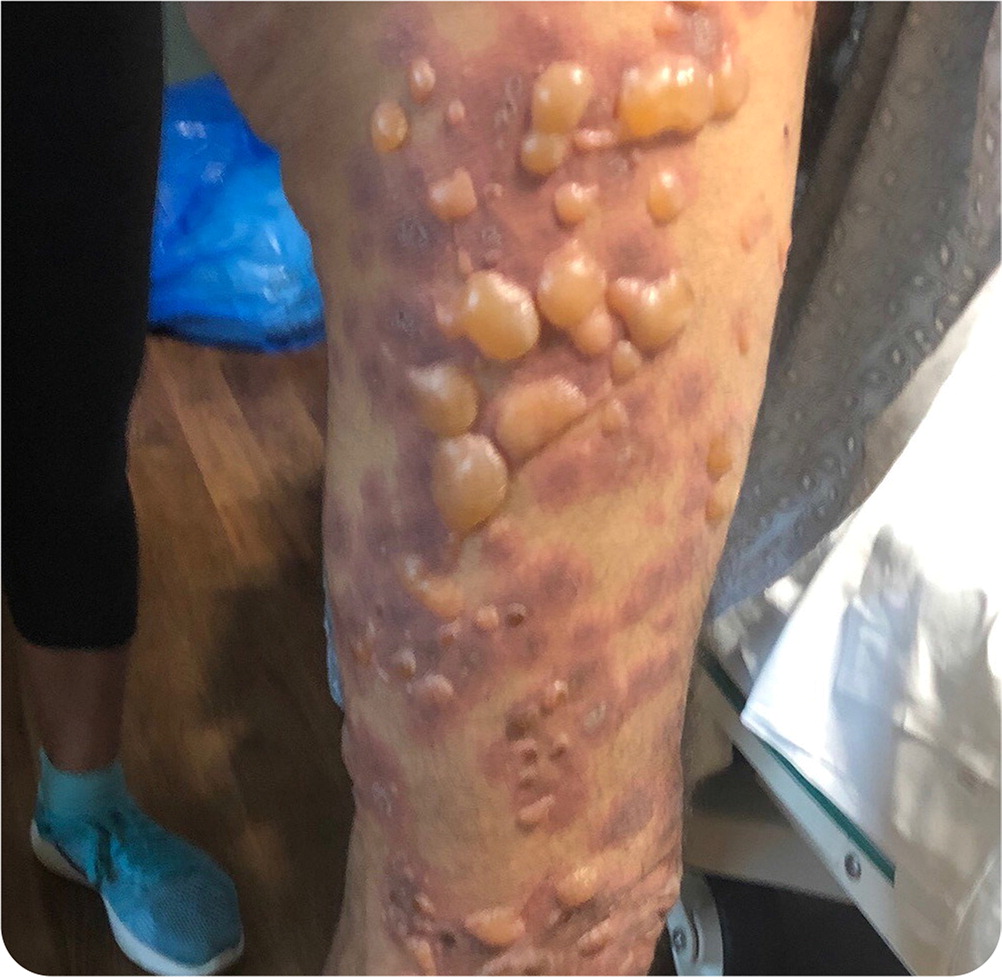
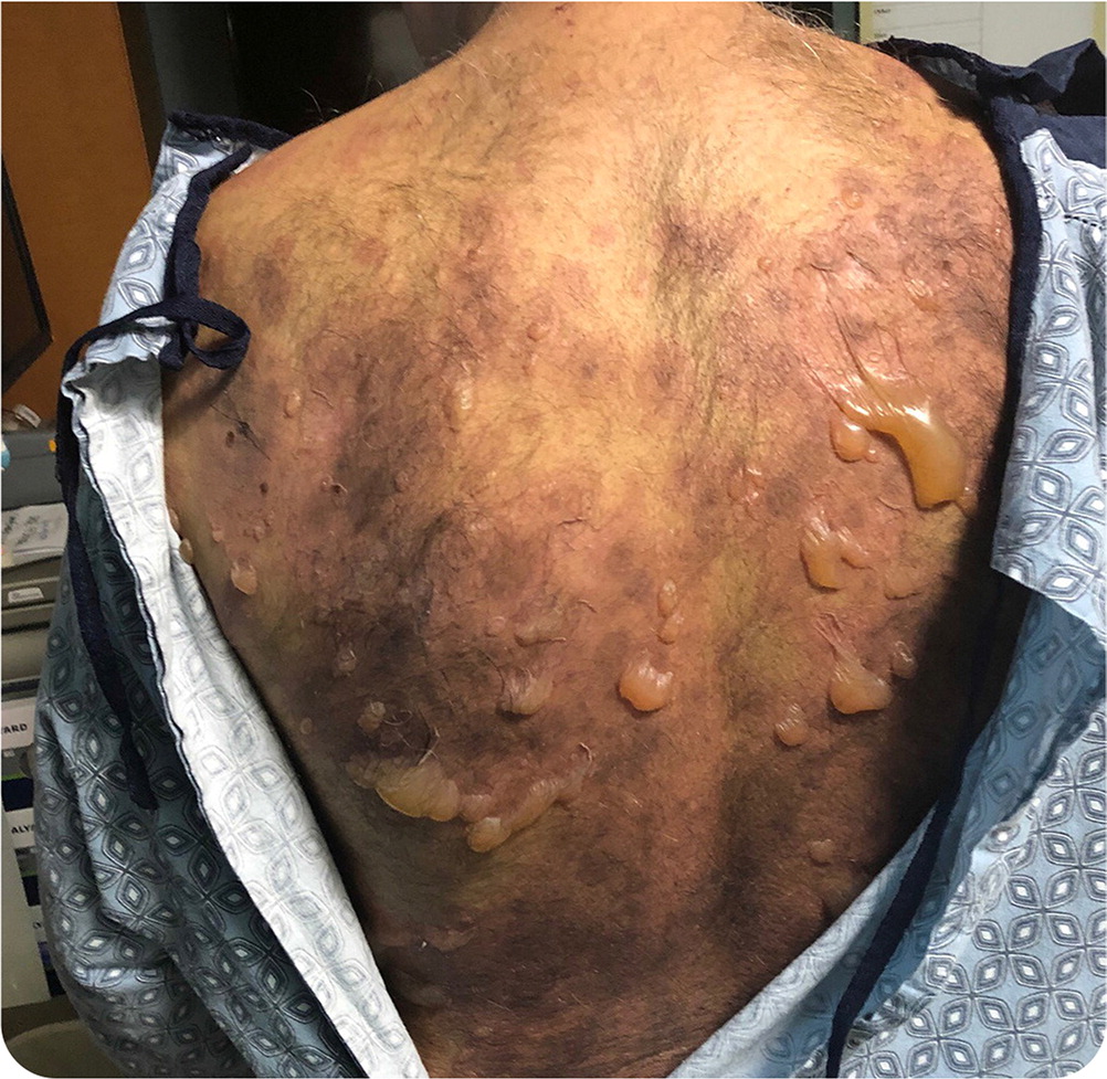
Physical examination revealed bullae the same color as the patient's skin on an erythematous base (Figure 1 and Figure 2). The bullae were in different stages, with some tense and filled with fluid and others flaccid and drained. The lesions varied in size from 1 to 10 cm and were located across the body, with the largest clusters on his arms, legs, chest, and back. No bullae were present on the patient's face, mouth, mucosa, or anal and genital areas. No excoriations were noted.


Question
Based on the patient's history and physical examination findings, which one of the following is the most likely diagnosis?
A. Bullous pemphigoid.
B. Staphylococcal scalded skin syndrome.
C. Stevens-Johnson syndrome.
D. Toxic epidermal necrolysis.
Discussion
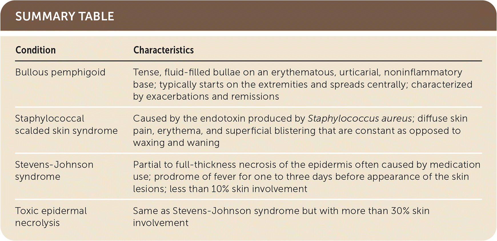
The answer is A: bullous pemphigoid, a chronic autoimmune skin condition that usually affects adults 60 to 80 years of age. It is characterized by exacerbations and remissions. Tense, fluid-filled bullae on an erythematous, urticarial, noninflammatory base typically start on the extremities and spread centrally. Mucosal involvement occurs in 10% to 30% of patients.1,2
Staphylococcal scalded skin syndrome is caused by the endotoxin produced by Staphylococcus aureus. It primarily occurs in childhood, although adults can also be affected. Patients present with diffuse skin pain, erythema, and superficial blistering that are constant as opposed to waxing and waning. Diagnosis is usually clinical; however, cultures of drainage fluid may show S. aureus. Treatment requires antibiotics and supportive measures.5
Stevens-Johnson syndrome and toxic epidermal necrolysis are similar life-threatening skin conditions. The main differentiating factor is the total body surface area involved. Stevens-Johnson syndrome is not as severe, with less than 10% skin involvement. In contrast, more than 30% of the total body surface area is involved with toxic epidermal necrolysis. Both conditions cause partial to full-thickness necrosis of the epidermis. These conditions are usually caused by medications. Both begin with a prodrome of fever for one to three days before skin lesions appear. Mucosal involvement occurs in 90% of patients with these conditions. The acute phase lasts eight to 12 days with persistent fevers, severe mucosal involvement, and epidermal sloughing.6
Treatment of Stevens-Johnson syndrome and toxic epidermal necrolysis is based on prompt cessation of the causative agent and appropriate in-patient placement (intensive care or burn unit) with supportive care, including wound care, intravenous fluids, and pain control.6

| Condition | Characteristics |
|---|---|
| Bullous pemphigoid | Tense, fluid-filled bullae on an erythematous, urticarial, noninflammatory base; typically starts on the extremities and spreads centrally; characterized by exacerbations and remissions |
| Staphylococcal scalded skin syndrome | Caused by the endotoxin produced by Staphylococcus aureus; diffuse skin pain, erythema, and superficial blistering that are constant as opposed to waxing and waning |
| Stevens-Johnson syndrome | Partial to full-thickness necrosis of the epidermis often caused by medication use; prodrome of fever for one to three days before appearance of the skin lesions; less than 10% skin involvement |
| Toxic epidermal necrolysis | Same as Stevens-Johnson syndrome but with more than 30% skin involvement |
