Am Fam Physician. 2023;107(1):87-88
Author disclosure: No relevant financial relationships.
A 36-year-old woman presented with a mass on her labia that she first noticed two years earlier. The mass had recently started to bleed, requiring multiple pad changes per day. The patient reported pain with walking and sitting. She did not have trauma or surgery in the affected area. She was obese (body mass index of 40 kg per m2) and had a history of irregular menses.
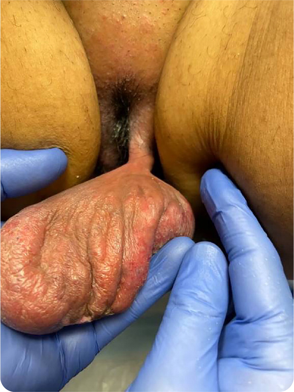
The physical examination revealed a large pedunculated mass stemming from the left labium majus. The gelatinous polyp measured approximately 13 × 8 × 4 cm with a 2- × 2-cm stalk (Figure 1). The surface had areas of ulceration, erosion, and scaling. The pelvic examination was otherwise unremarkable. Ultrasonography revealed an echogenic, irregularly shaped lesion with vascularization and no evidence of peristalsis.

Question
Based on the patient's history and physical examination findings, which one of the following is the most likely diagnosis?
A. Bartholin gland cyst.
B. Canal of Nuck cyst.
C. Fibroepithelial stromal polyp.
D. Gartner duct cyst.
E. Neurofibroma.
Discussion
The answer is C: fibroepithelial stromal polyp of the vulva. This is a benign lesion of mesenchymal origin in women of childbearing age. It is typically characterized by a solitary lesion the same color as the patient's skin, and it is attached by a long stalk. Fibroepithelial polyps are uncommon, hormone-sensitive masses that usually appear in the vagina, although they can occur in the cervix and vulva. Polyps up to 20 cm in size have been described in rare case reports. Although asymptomatic, they can present with discharge and bleeding. The patient's solitary, irregularly shaped, pedunculated lesion with areas of ulceration and erosion is characteristic of a fibroepithelial polyp. Histologic evaluation can rule out malignancy. Treatment consists of surgical excision, and recurrence is rare.1
The Bartholin glands are located in the labia minora and drain through ducts in the posterior vagina. The glands secrete vaginal lubricating mucus. Bartholin ducts can become obstructed, causing a cyst. An abscess may form if they become infected. These lesions are typically unilateral and present as swelling and tenderness in the posterior vulva. They may be tender or red. They are not typically pedunculated.2
A canal of Nuck cyst is a hydrocele that protrudes through the inguinal canal. The canal of Nuck is an area anterior to the round ligament that protrudes through the inguinal ring toward the labia majora. The canal of Nuck usually resolves before birth but, if persisting, can lead to an inguinal hernia or a hydrocele. The cyst presents as a translucent lump on the labia, is worse with standing, and improves when supine. On ultrasonography, it appears as a thin-walled cystic structure. Rarely, bowel can herniate into the mass. It is diagnosed through surgical exploration.3
A Gartner duct is a remnant of the mesonephric (wolffian) duct that normally disappears during embryogenesis but occasionally remains and forms a vaginal inclusion cyst. The Gartner ducts, located on either side of the urethra, are often associated with other renal anomalies. Gartner duct cysts typically develop on the anterior and lateral parts of the vaginal wall, but if they are large, they may appear midline in the vagina.4
A neurofibroma is a benign peripheral nerve sheath tumor that can occur anywhere on the skin, most commonly the abdomen, chest, and back. It can be solitary or multiple, as with neurofibromatosis. There have been case reports of large neurofibromas on the labia. A neurofibroma typically appears as a small, firm, rubbery subcutaneous nodule or papule that can become pedunculated.5

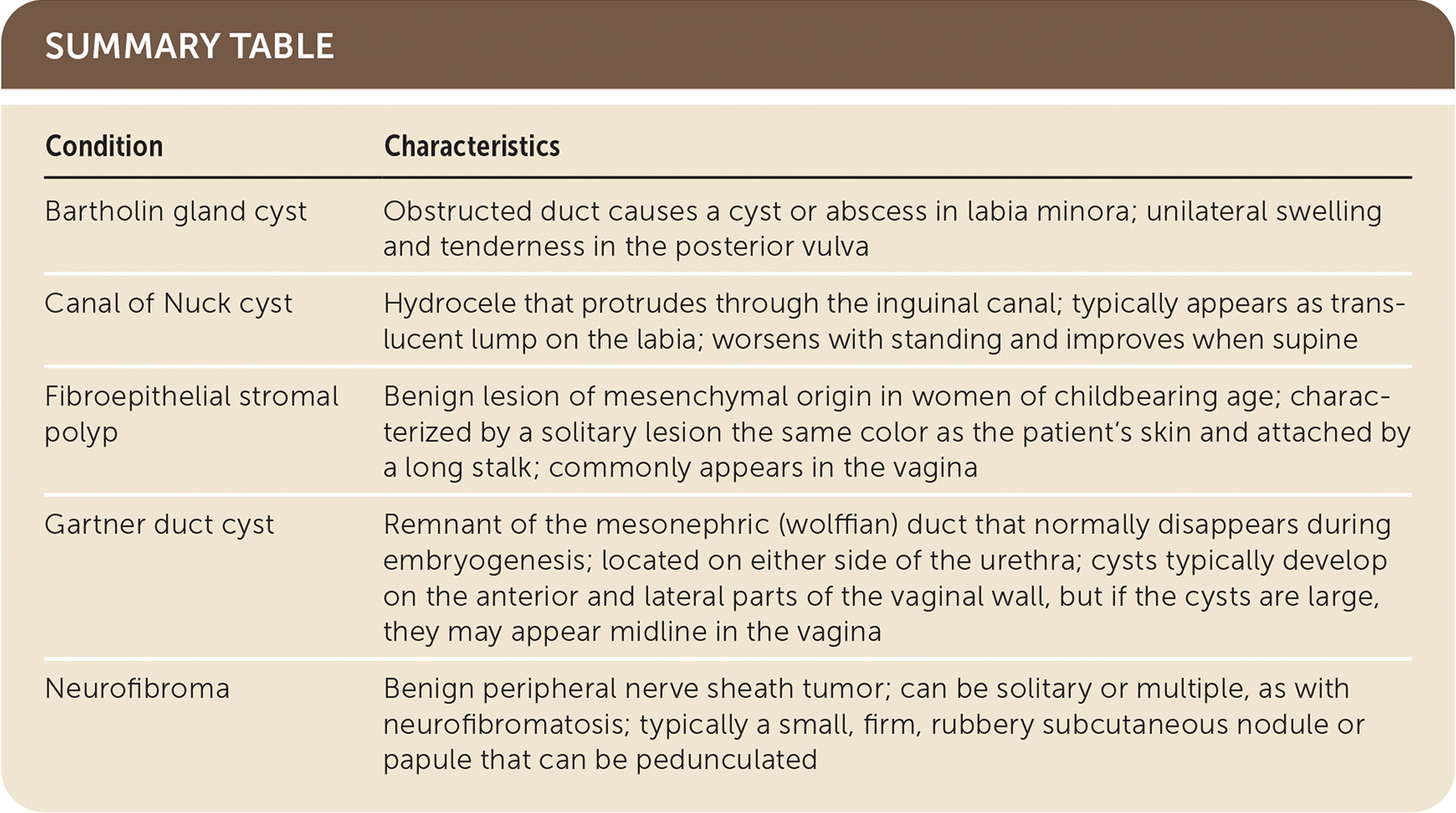
| Condition | Characteristics |
|---|---|
| Bartholin gland cyst | Obstructed duct causes a cyst or abscess in labia minora; unilateral swelling and tenderness in the posterior vulva |
| Canal of Nuck cyst | Hydrocele that protrudes through the inguinal canal; typically appears as translucent lump on the labia; worsens with standing and improves when supine |
| Fibroepithelial stromal polyp | Benign lesion of mesenchymal origin in women of childbearing age; characterized by a solitary lesion the same color as the patient's skin and attached by a long stalk; commonly appears in the vagina |
| Gartner duct cyst | Remnant of the mesonephric (wolffian) duct that normally disappears during embryogenesis; located on either side of the urethra; cysts typically develop on the anterior and lateral parts of the vaginal wall, but if the cysts are large, they may appear midline in the vagina |
| Neurofibroma | Benign peripheral nerve sheath tumor; can be solitary or multiple, as with neurofibromatosis; typically a small, firm, rubbery subcutaneous nodule or papule that can be pedunculated |
