
Am Fam Physician. 2023;107(2):191-192
Author disclosure: No relevant financial relationships.
A 58-year-old woman with no significant medical history presented with well-demarcated, violaceous nodules on her face (Figure 1) and dusky, erythematous plaques on her legs (Figure 2). The lesions appeared five months earlier. They were asymptomatic but rapidly growing. The patient reported fatigue and malaise.


Biopsy and blood tests were performed. Punch biopsy of the left thigh revealed irregular vascular spaces, endothelial atypia, and increased spindle cells with numerous plasma cells (Figure 3).

Question
Based on the patient's history, physical examination, and biopsy findings, which one of the following is the most likely diagnosis?
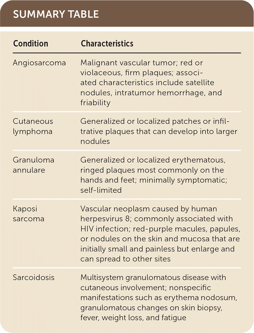
A. Angiosarcoma.
B. Cutaneous lymphoma.
C. Granuloma annulare.
D. Kaposi sarcoma.
E. Sarcoidosis.
Discussion
The answer is D: Kaposi sarcoma, a vascular neoplasm caused by human herpesvirus 8. It is commonly associated with HIV infection. Kaposi sarcoma presents as red-purple macules, papules, or nodules on the skin and mucosa. There are several stages of Kaposi sarcoma, including the patch, plaque, and tumor stages. Although initially small and painless, lesions enlarge and spread. Histopathologic findings include increased spindle cells and vascular structures in the dermis, with prominent plasma cell infiltration.1
When associated with HIV, Kaposi sarcoma is an AIDS-defining illness, although it is becoming less common in the United States due to the use of highly active antiretroviral therapy. Kaposi sarcoma should be considered in patients with unknown HIV status. This patient subsequently tested positive for HIV.
Angiosarcoma is a malignant vascular tumor commonly found on the head and neck in older people, areas of chronic lymphedema, or previously irradiated areas. Lesions present as red or violaceous, firm plaques with associated characteristics such as satellite nodules, intratumor hemorrhage, and friability. Prognosis is poor for all subtypes. On histopathology, atypical endothelial cells line vascular spaces in the dermis.2
Cutaneous lymphoma can present as a primary manifestation or secondary to an underlying systemic lymphoma. Most cutaneous lymphomas originate with the T cells and present as generalized or localized patches or infiltrative plaques in the early stages and as larger nodules at the tumor stage.3 On histopathology, lymphocytes are large and dark with irregular nuclear contours and line the dermal-epidermal junction. Vacuolar interface changes are also present.4
Granuloma annulare presents as erythematous, ringed plaques most commonly on the hands and feet. Clinical variants include generalized, localized, and subcutaneous subtypes. The etiology is often unknown, and lesions are minimally symptomatic and self-limited.5 Granuloma annulare can have interstitial or palisading patterns of granulomatous inflammation involving the upper to mid-reticular dermis.6
Sarcoidosis is a multisystem granulomatous disease, with cutaneous involvement in approximately 40% of cases. There are many nonspecific manifestations such as erythema nodosum, granulomatous changes on skin biopsy, fever, weight loss, and fatigue. Lung involvement and lymphadenopathy should raise suspicion for sarcoidosis.7 On histopathology, sarcoidosis presents as epithelioid histiocytes with minimal inflammation.6
| Condition | Characteristics |
|---|---|
| Angiosarcoma | Malignant vascular tumor; red or violaceous, firm plaques; associated characteristics include satellite nodules, intratumor hemorrhage, and friability |
| Cutaneous lymphoma | Generalized or localized patches or infiltrative plaques that can develop into larger nodules |
| Granuloma annulare | Generalized or localized erythematous, ringed plaques most commonly on the hands and feet; minimally symptomatic; self-limited |
| Kaposi sarcoma | Vascular neoplasm caused by human herpesvirus 8; commonly associated with HIV infection; red-purple macules, papules, or nodules on the skin and mucosa that are initially small and painless but enlarge and can spread to other sites |
| Sarcoidosis | Multisystem granulomatous disease with cutaneous involvement; nonspecific manifestations such as erythema nodosum, granulomatous changes on skin biopsy, fever, weight loss, and fatigue |
