
Am Fam Physician. 2023;107(2):204-206
Author disclosure: No relevant financial relationships.
Key Points for Practice
• Attempt to perform 12-lead ECG in patients with chest pain within 10 minutes of arrival to a clinic or emergency setting.
• Use a clinical decision pathway to identify patients with low-risk chest pain who can be discharged from the emergency department.
• If available, use CCTA preferentially over stress testing for patients with intermediate-risk chest pain to determine the need for invasive coronary angiography. For patients with high-risk chest pain, provide referral for invasive coronary angiography.
• In patients with known CAD, focus on controlling blood pressure and cholesterol. Consider CCTA to document CAD progression in patients with previous testing demonstrating nonobstructive lesions.
From the AFP Editors
Chest pain leads to about 4 million outpatient visits per year and is the second most common reason for emergency department care, with nearly 7 million visits per year. Although most chest pain is noncardiac, more than 18 million people in the United States have coronary artery disease (CAD), leading to more than 1,000 deaths per day. The American Heart Association/American College of Cardiology (AHA/ACC) updated guidelines for management of chest pain, which are endorsed by five other cardiology groups. The guidelines provide new recommendations on what to consider chest pain and when to avoid testing in patients at low risk, and they endorse use of published decision pathways to determine the order and extent of workup.
Initial Evaluation
Chest pain can present as pain, pressure, tightness, or discomfort in the chest, shoulders, arms, neck, back, upper abdomen, or jaw and less commonly as shortness of breath, nausea, or fatigue without pain. Chest pain is considered acute with new onset or if it involves a change in pattern, intensity, or duration; it is considered stable if it is chronic with unchanging triggers such as exertion or emotional stress. Patients commonly describe ischemic chest pain as pressure, squeezing, heaviness, tightness, exertional, stress-related, or retrosternal. Pain that is sharp, fleeting, pleuritic, positional, or shifting locations is less likely to be of cardiac origin. These guidelines suggest describing chest pain as cardiac; possible cardiac; or noncardiac. The descriptor atypical is no longer used because it can be interpreted by patients as being benign.
Initial evaluation should focus on ruling out life-threatening illnesses such as acute coronary syndrome (ACS), aortic dissection, and pulmonary embolism, which are often not indicated by pain severity. Response to nitroglycerin is not an accurate means of ruling in cardiac chest pain. Patients with diabetes mellitus, women, and older patients more often present with associated nausea, fatigue, and shortness of breath. ACS should be considered when patients older than 75 years present with shortness of breath, syncope, mental impairment, or abdominal pain, or if they experience an unexplained fall.
People from ethnic and racial minorities experience delays in diagnosis of a cardiac cause and treatment of chest pain. A higher proportion of Black patients presenting with chest pain have a cardiac etiology, but they are less likely to receive appropriate diagnostic testing and urgent treatment. Treatment disparities are also found in Hispanic and South Asian patients and in people who are uninsured or are insured by Medicaid.
Diagnostic Evaluation
Initial evaluation should involve 12-lead electrocardiography (ECG), which is recommended within 10 minutes of arrival in clinic and emergency settings. Because up to 6% of patients with cardiac ischemia are discharged from the emergency department after a single normal ECG, repeat testing should be considered in patients with normal ECG but a higher index of suspicion. Adding leads V7 to V9 should be considered on repeat testing to assess for posterior wall ischemia. ST elevation, hyperacute T waves, left bundle branch block, and ST depression are the most concerning findings for cardiac chest pain.
Physical examination can sometimes demonstrate life-threatening causes of chest pain, including diaphoresis and tachypnea suggesting ACS, tachycardia and dyspnea suggesting pulmonary embolism, and subcutaneous emphysema suggesting esophageal rupture. Chest tenderness on palpation or pain with inspiration suggests a noncardiac etiology.
Radiography can demonstrate chest pain etiologies such as pneumonia, pneumothorax, and rib fracture. Although it may suggest aortic dissection, lack of a widened mediastinum cannot rule out dissection. Prompt transthoracic echocardiography is recommended, if available, for rapid evaluation of cardiac function.
High-sensitivity cardiac troponin is the most accurate and early marker of cardiac injury. The creatine kinase myocardial isoenzyme and myoglobin are not useful for myocardial injury diagnosis or prognosis.
Risk Stratification Testing
Cardiac computed tomographic angiography (CCTA) can be used to identify obstructive CAD that increases the risk of major coronary events in patients younger than 65 years or without known obstructive CAD. Stress testing should be considered for patients older than 65 years, those with inconclusive anatomic studies, or when more obstructive CAD is suspected.
Anatomic testing with CCTA and invasive coronary angiography demonstrates the extent of obstructive disease. Adding a calculation of fractional flow reserve with computed tomography to CCTA provides an estimation of lesion-specific ischemia. Invasive coronary angiography identifies obstructive stenosis and allows revascularization. CCTA has an effective dose of 3 to 5 mSv, compared with 4 to 10 mSv for angiography. CCTA without stenosis or plaque has a warranty period of two years for similar symptom frequency and negative troponin testing.
Stress imaging can include exercise or pharmacologic stress and assessment with ECG, echocardiography, nuclear imaging with positron emission tomography, single photon emission computed tomography, or cardiovascular magnetic resonance imaging (MRI). Positron emission tomography is more accurate than single photon emission computed tomography and should be used if available. Cardiovascular MRI can also accurately assess global and regional left and right ventricular function, detect and localize myocardial ischemia and infarction, and determine myocardial viability in patients not able to exercise. A normal stress test has a warranty period of one year for similar symptom frequency and negative troponin testing.
Exercise can be used for any patient who is not frail and can achieve at least five metabolic equivalents, which is required to do many activities of daily living. Patients who cannot meet both requirements should receive pharmacologic stress testing. Exercise ECG testing should be avoided in patients with ECG findings of 0.5-mm ST depression, left ventricular hypertrophy, paced rhythm, left bundle branch block, Wolff-Parkinson-White pattern, or digitalis use. Exercise ECG testing is equally predictive of future events as stress imaging, despite lower sensitivity for obstruction. Failure to exceed five metabolic equivalents or to achieve 85% of predicted heart rate suggests a poor prognosis, whereas exceeding 10 metabolic equivalents confers a low risk of cardiac events. Positive exercise ECG results can be further clarified by CCTA or functional fractional reserve testing.
All risk stratification modalities have a low radiation risk to the fetus and can be used in pregnant patients. Iodinated contrast enters fetal circulation and should be used with caution in pregnant patients. Patients can continue to breastfeed because only 1% of iodinated contrast is excreted into breast milk and absorbed. Gadolinium contrast in cardiovascular MRI should be used only when necessary.
Clinical Decision Pathways
Decision pathways based on high-sensitivity troponin levels, such as the HEART Pathway, EDACS, mADAPT, NOTR, or the 2020 European Society of Cardiology pathway, should be used to determine the need for testing and hospitalization. Using these pathways can decrease hospital admission and unnecessary testing by up to 43%. A second high-sensitivity troponin test should be ordered one to three hours following the first.
Low-risk patients with acute chest pain do not benefit from stress testing or cardiac imaging within 30 days of initial visit. Coronary artery calcium scoring is not routinely needed; a score of 0 denotes low risk, although positive scores do not reliably identify patients at increased risk.
For patients without known CAD at intermediate risk, CCTA is recommended with a negative or inconclusive ACS workup and for patients with a mildly abnormal stress test within the past year. CCTA allows for rapid diagnosis and discharge with similar outcomes as stress testing. CCTA decreases time to diagnosis by 50% compared with nuclear stress testing. Negative stress testing rules out the need for anatomic testing. Exercise ECG testing and stress echocardiography are most likely to reduce length of stay.
Intermediate-risk patients with known CAD and acute chest pain should have blood pressure and cholesterol management maximized before additional testing. CCTA can document CAD progression in patients with previous testing demonstrating nonobstructive lesions. Patients with a history of high-risk CAD or worsening frequency of symptoms who have optimized blood pressure and cholesterol management should be evaluated with invasive coronary angiography.
High-risk patients with acute chest pain should receive invasive coronary angiography.
Evaluation of Patients With Stable Chest Pain
In patients with stable chest pain, the risk algorithm from the CAD Consortium (https://reference.medscape.com/calculator/287/pre-test-probability-of-cad-cad-consortium) guides testing. Low-risk patients will not need further testing.
For patients at intermediate or high risk, CCTA is preferred; stress testing involving echocardiography or nuclear testing can be used instead of cardiovascular MRI.
Stress testing should be considered in patients with known obstructive CAD and continuing stable chest pain only if blood pressure and cholesterol management is optimized. Invasive coronary angiography should be considered to help guide pharmacologic therapy in patients with a history of coronary artery bypass grafting and concern for myocardial ischemia and in patients with indeterminate or nondiagnostic stress test results.
In patients with suspected ischemia despite no obstructive cardiac disease, positron emission tomography, cardiovascular MRI, or angiography may help tailor medical therapy to improve quality of life, although they have not been demonstrated to reduce future cardiac events.
Nonischemic Chest Pain
Immediate echocardiography should be used in patients with suspected nonischemic causes of chest pain such as aortic dissection, pericardial effusion, and pulmonary embolism. If echocardiography rules out life-threatening injury, computed tomography or cardiovascular MRI can be used to confirm a suspected diagnosis.
In patients with nonischemic causes of chest pain, gastrointestinal causes, depression, and anxiety should be considered. Up to one-fifth of people presenting with chest pain have a gastrointestinal cause. In patients with low-risk chest pain, depression, anxiety, and gastrointestinal disease each exceed CAD by a factor of 10. Although mental health referrals are rare even when patients report anxiety, psychotherapy for these patients reduces chest pain frequency by one-third.
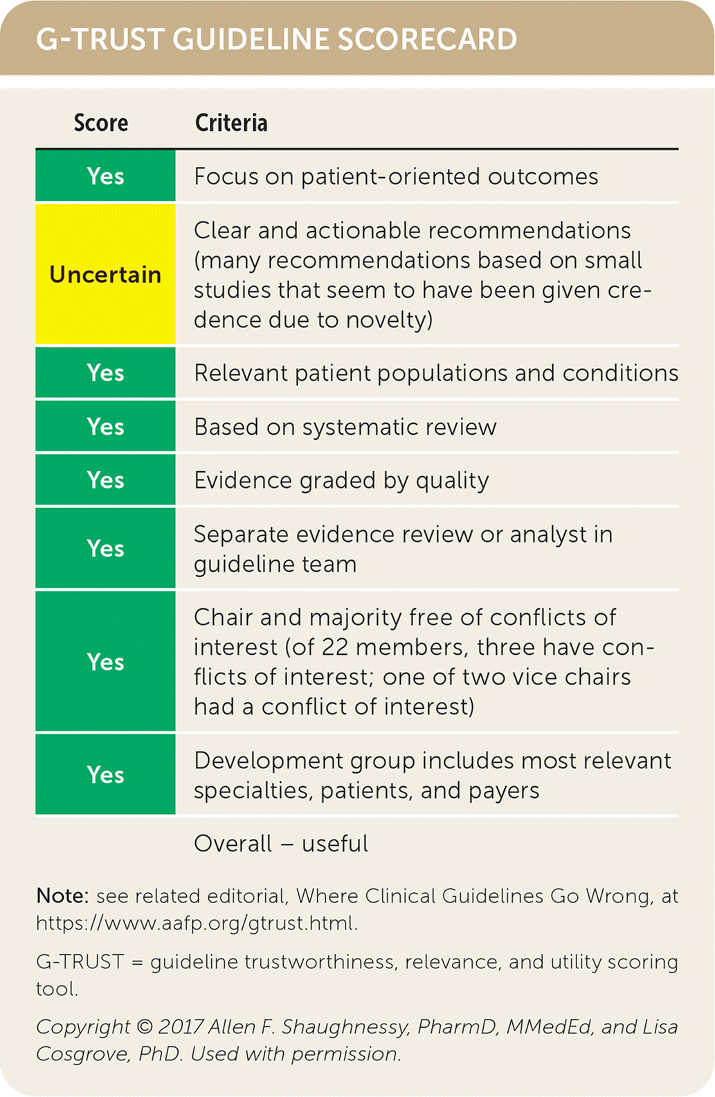
| Score | Criteria |
|---|---|
| Yes | Focus on patient-oriented outcomes |
| Uncertain | Clear and actionable recommendations (many recommendations based on small studies that seem to have been given credence due to novelty) |
| Yes | Relevant patient populations and conditions |
| Yes | Based on systematic review |
| Yes | Evidence graded by quality |
| Yes | Separate evidence review or analyst in guideline team |
| Yes | Chair and majority free of conflicts of interest (of 22 members, three have conflicts of interest; one of two vice chairs had a conflict of interest) |
| Yes | Development group includes most relevant specialties, patients, and payers |
| Overall – useful |
Guideline source: American Heart Association/American College of Cardiology
Published source: J Cardiovasc Comput Tomogr. January/February 2022;16(1):54–122
Editor's Note: These guidelines are an important update because they clarify risk stratification of chest pain in two ways. First, AHA/ACC recommends that testing be guided by cardiac risk using one of the many clinical decision pathways, which are simple risk algorithms that use high-sensitivity troponin and clinical characteristics. If you do not have a favorite risk algorithm, the HEART pathway is easy to use and is available through MDCalc (https://www.mdcalc.com/calc/3975/heart-pathway-early-discharge-acute-chest-pain). It optimizes the accuracy of identifying more patients at low risk. Also, these guidelines clarify that CCTA should be our default risk stratification method for intermediate-risk patients with acute chest pain because it saves time compared with nuclear stress testing with lower cost and radiation exposure compared with invasive angiography.—Michael J. Arnold, MD, Contributing Editor
The views expressed are those of the authors and do not necessarily reflect the official policy or position of the U.S. Department of the Navy, Uniformed Services University of the Health Sciences, U.S. Department of Defense, U.S. Department of Veterans Affairs, or the U.S. government.
